

Review
doi: 10.1007/s10974-013-9355-8.
Epub 2013 Aug 1.
Cytoskeletal tropomyosins: choreographers of actin filament functional diversity
Affiliations
- PMID: 23904035
- PMCID: PMC3843815
- DOI: 10.1007/s10974-013-9355-8
Item in Clipboard
Review
Cytoskeletal tropomyosins: choreographers of actin filament functional diversity
J Muscle Res Cell Motil.
2013 Aug.
Abstract
The actin cytoskeleton plays a central role in many essential cellular processes. Its involvement requires actin filaments to form multiple populations with different structural and therefore functional properties in specific subcellular locations. This diversity is facilitated through the interaction between actin and a number of actin binding proteins. One family of proteins, the tropomyosins, are absolutely essential in regulating actin's ability to form such diverse structures. In this review we integrate studies from different organisms and cell types in an attempt to provide a unifying view of tropomyosin dependent regulation of the actin cytoskeleton.
Figures
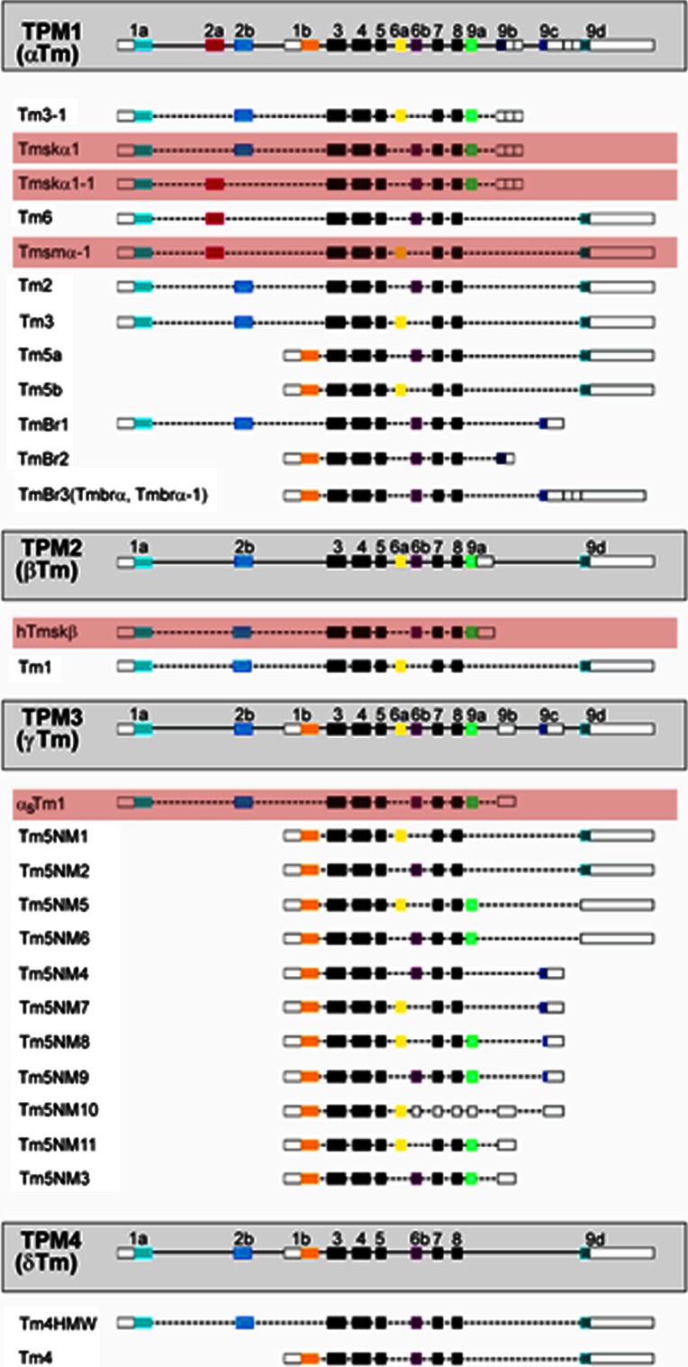
Diagram of the TPM1 (α), TPM2 (β), TPM3 (γ) and TPM4 (δ) genes and the isoforms they encode. The white boxes show untranslated regions, dotted lines represent introns and the black boxes show exons common to all isoforms. Muscle isoforms (shown highlighted in red) account for only five tropomyosin isoforms expressed in mammalian cells. Only the major isoforms are included, a number of mRNAs have been detected only by RT-PCR and are not shown
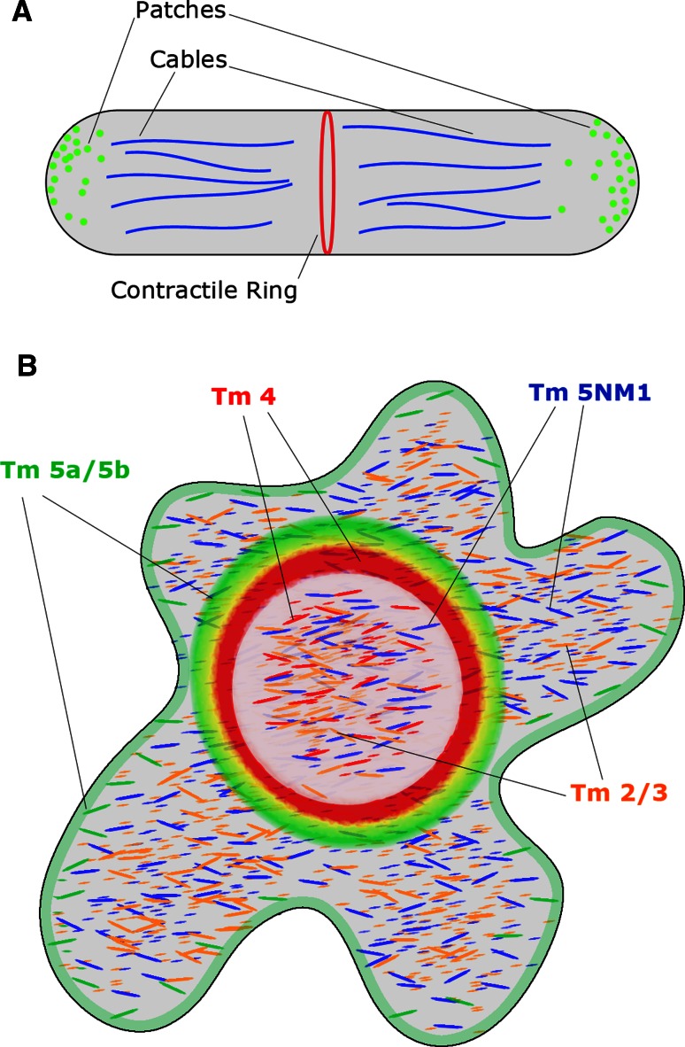
a Distribution of tropomyosin in Schizosaccharomyces pombe. Actin patches are found near the cell periphery and are not associated with tropomyosin isoforms. The cables which run throughout the cell are associated with unacetylated Cdc8p and favour the binding of myosin-V. In contrast, the contractile ring actin is associated with acetylated Cdc8p which favours the binding of myosin-II. b Distribution of tropomyosin in Osteoclasts plated on ivory. Tm4 (red) is associated with podosomes (represented as the inner ring) and the interior of the cell. Tm5a/5b (green) is associated with the F-actin ring (represented as the outer ring) and is slightly enriched near the plasma membrane. Whilst some colocalization is observed between Tm5a/5b and Tm4 (yellow), Tm5a/5b are notably absent from the podosomes. Both Tm2/3 (orange) and Tm5NM1 (blue) are both found throughout the cell in different subcellular pools, however the nature of these regions is not yet known
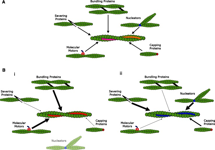
a Previous view: Actin filaments are formed independent of any association with actin binding proteins aside from their nucleators. Once formed tropomyosin isoforms nonspecifically dimerize with any isoform of the same molecular weight. At a critical concentration these homo- or heterodimers will form heteropolymers along the filament where they act only to stabilize the actin filament. b Current model: Actin filaments are nucleated and begin to polymerize. Specific tropomyosin isoforms co-polymerize with the newly nucleated filaments stabilizing them whilst polymerization continues. Once the mature filament has formed the bound tropomyosin regulates the interaction between actin and actin binding proteins. (i) Seen above the red decorated filaments there is enhanced binding of bundling proteins and molecular motors, whilst nucleators and capping protein interaction is greatly diminished. There is also complete exclusion of severing proteins from these structures. (ii) In contrast, filaments decorated by blue tropomyosin enhance the severing protein’s binding whilst modulating the access of other actin binding proteins independent of the red filaments. This allows for the formation of multiple filament populations which are able to perform specific cellular functions and that are independently regulated in different regions of the cell
References
-
- Bryce NS, Schevzov G, Ferguson V, Percival JM, Lin JJ-C, Matsumura F, Bamburg JR, Jeffrey PL, Hardeman EC, Gunning P, Weinberger RP. Specification of actin filament function and molecular composition by tropomyosin isoforms. Mol Biol Cell. 2003;14(3):1002–1016. doi: 10.1091/mbc.E02-04-0244. - DOI - PMC - PubMed
Publication types
MeSH terms
Substances
LinkOut - more resources
Full Text Sources
Other Literature Sources
