. 2013 Oct;22(10):1336-48.
doi: 10.1002/pro.2320. Epub 2013 Aug 19.

Nonuniform chain collapse during early stages of staphylococcal nuclease folding detected by fluorescence resonance energy transfer and ultrarapid mixing methods

Affiliations

Nonuniform chain collapse during early stages of staphylococcal nuclease folding detected by fluorescence resonance energy transfer and ultrarapid mixing methods

Takuya Mizukami et al. Protein Sci. 2013 Oct.

Abstract

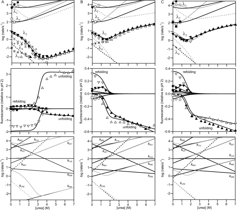
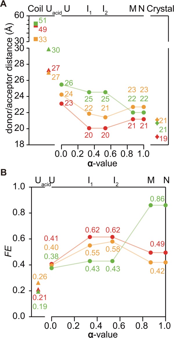
The development of tertiary structure during folding of staphylococcal nuclease (SNase) was studied by time-resolved fluorescence resonance energy transfer measured using continuous- and stopped-flow techniques. Variants of this two-domain protein containing intradomain and interdomain fluorescence donor/acceptor pairs (Trp and Cys-linked fluorophore or quencher) were prepared to probe the intradomain and interdomain structural evolution accompanying SNase folding. The intra-domain donor/acceptor pairs are within the β-barrel domain (Trp27/Cys64 and Trp27/Cys97) and the interdomain pair is between the α-helical domain and the β-barrel domain (Trp140/Cys64). Time-resolved energy transfer efficiency accompanying folding and unfolding at different urea concentrations was measured over a time range from 30 μs to ≈ 10 s. Information on average donor/acceptor distances at different stages of the folding process was obtained by using a quantitative kinetic modeling approach. The average distance for the donor/acceptor pairs in the β-barrel domain decreases to nearly native values whereas that of the interdomain donor/acceptor pairs remains unchanged in the earliest intermediate (<500 μs of refolding). This indicates a rapid nonuniform collapse resulting in an ensemble of heterogeneous conformations in which the central region of the β-barrel domain is well developed while the C-terminal α-helical domain remains disordered. The distance between Trp140 and Cys64 decreases to native values on the 100-ms time scale, indicating that the α-helical domain docks onto the preformed β-barrel at a late stage of the folding. In addition, the unfolded state is found to be more compact under native conditions, suggesting that changes in solvent conditions may induce a nonspecific hydrophobic collapse.

Keywords: Förster energy transfer; continuous-flow; fluorescence; protein folding; stopped-flow.


Figures

Scheme 1
Scheme 1
Kinetic mechanism of folding of SNase.
Figure 1
Figure 1
Ribbon diagram of Trp140 SNase (P47G/P117G/H124L) based on the crystal structure (PDB 1SNQ). The Cβ atoms of Tyr27, Trp140, Lys64, and Lys97 are shown as spheres. The Cβ-Cβ distances of Tyr27-Lys64, Tyr27-Lys97, and Trp140-Lys64 are shown in dotted lines. The β-barrel domain is encircled.
Figure 2
Figure 2
Absorption spectra of the dye-labeled proteins and Trp fluorescence spectra of the unmodified proteins. Absorption spectra of (A) Trp140/Cys64-TNB, (B) Trp27/Cys64-AED, and (C) Trp27/Cys97-AED (at 10 μM with a pathlength of 1 cm) under the native condition (∼100 mM sodium acetate at pH 5.2; black dashed lines) and the acid-denaturing condition (∼20 mM phosphoric acid at pH 2.0; gray solid lines), and Trp fluorescence spectra (dotted lines) of (A) Trp140/Cys64 SNase, (B) Trp27/Cys64 SNase, and (C) Trp27/Cys97 SNase (at 10 μM), under the native (black dotted lines) and the acid-denaturing conditions (gray dotted lines) at 15°C.
Figure 3
Figure 3
Time dependence of fluorescence intensity and the energy transfer efficiency during the folding reactions of (A) Trp140/Cys64, (B) Trp27/Cys64, and (C) Trp27/Cys97 variants initiated by a pH-jump from 2.0 to 5.2 (∼100 mM sodium acetate at pH 5.2) at 15°C. The upper panels show the kinetic traces of the pH-induced folding reaction of the unmodified (D) and the dye-labeled (D&A) proteins. The extrapolated fluorescence intensities of unfolded states at 0 M urea based on equilibrium measurements for each variant are shown in circles. The solid lines show the kinetic traces reproduced by the kinetic modeling. Fluorescence was monitored with a combination of a 305-nm long-pass filter and a 278/366-nm band-pass filter. The lower panels show the energy transfer efficiency (FRET) during folding calculated by Eq. (4).
Figure 4
Figure 4
Urea-dependence of the rate constants (chevron plot; upper panels), the kinetic amplitudes (middle panels) and the elementary rate constants obtained by kinetic modeling based on Scheme 1 (lower panels) for the unmodified and the dye-labeled (A) Trp140/Cys64, (B) Trp27/Cys64, and (C) Trp27/Cys97 variants. The refolding and unfolding measurements were carried out in ∼100 mM sodium acetate at pH 5.2 and 15°C. Upper and middle panels: The open and filled symbols show the rate constants and the kinetic amplitudes of the unmodified and the dye-labeled proteins, respectively, on the major folding pathway in Scheme 1. Circles, squares, and lower triangles represent the λ1, λ2, and λ3 phases of the folding, respectively, whereas upper triangles represent the λ3 phase of the unfolding. The corresponding rate constants and kinetic amplitudes are reproduced by the kinetic modeling and shown in black solid lines. Upper panels: The + and # symbols and the × symbols show the rate constants of the unmodified and the dye-labeled proteins, respectively, on the minor folding pathways (including the phase rate-limited by the peptidyl prolyl isomerization). The rate constants reproduced by the kinetic modeling are shown in black dashed lines. Previous kinetic modeling results on Trp140 SNase (A) and Trp76 SNase (B and C) are also shown in gray lines. Lower panels: The elementary rate constants (black lines; Supporting Information Table S1) reproducing the rate constants (gray lines) are shown.
Figure 5
Figure 5
(A) donor/acceptor distances of Trp140/Cys64-TNB (green, upper), Trp27/Cys64-AED (orange, middle), and Trp27/Cys97-AED (red, lower) in each state. Squares show the distances in random coil, estimated according to Refs. 37 and 38. Triangles show the distances in the acid unfolded state. Circles show the distance changes during folding from U to N states as a function of the α-value. Diamonds show Cβ-Cβ distance of the sites in the crystal structure. (B) FRET efficiency of Trp140/Cys64-TNB (green, upper), Trp27/Cys64-AED (orange, middle), and Trp27/Cys97-AED (red, lower) in each state. Triangles show FE in the acid unfolded state. Circles show the changes in FE during folding from U to N states as a function of the α-value.
Figure 6
Figure 6
Equilibrium unfolding of the unmodified (open symbols) and the dye-labeled (filled symbols) (A) Trp140/Cys64, (B) Trp27/Cys64, and (C) Trp27/Cys97 variants under matching condition of the kinetic measurements (∼100 mM sodium acetate at pH 5.2 and 15°C). Also shown are final fluorescence intensities of (un)folding kinetics in circles, initial fluorescence intensities of folding kinetics in squares, and initial fluorescence intensities of unfolding kinetics in diamonds.

References

    1. Baldwin RL, Rose GD. Is protein folding hierarchic? II. Folding intermediates and transition states. Trends Biochem Sci. 1999;24:77–83. - PubMed
    1. Arai M, Kuwajima K. Role of the molten globule state in protein folding. Adv Protein Chem. 2000;53:209–282. - PubMed
    1. Roder H, Maki K, Cheng H. Early events in protein folding explored by rapid mixing methods. Chem Rev. 2006;106:1836–1861. - PMC - PubMed
    1. Udgaonkar JB, Baldwin RL. NMR evidence for an early framework intermediate on the folding pathway of ribonuclease A. Nature. 1988;335:694–699. - PubMed
    1. Roder H, Wüthrich K. Protein folding kinetics by combined use of rapid mixing techniques and NMR observation of individual amide protons. Proteins. 1986;1:34–42. - PubMed

LinkOut - more resources