. 2013 Jul 31;33(31):12679-88.
doi: 10.1523/JNEUROSCI.0645-13.2013.

Anterobasal temporal lobe lesions alter recurrent functional connectivity within the ventral pathway during naming

Affiliations

Anterobasal temporal lobe lesions alter recurrent functional connectivity within the ventral pathway during naming

Pablo Campo et al. J Neurosci. .

Abstract

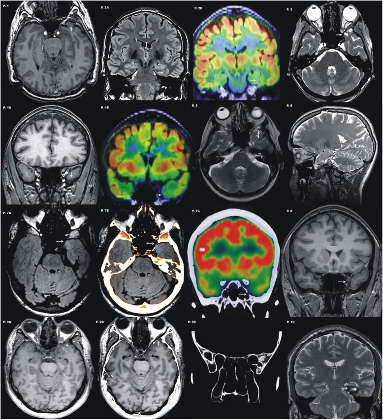
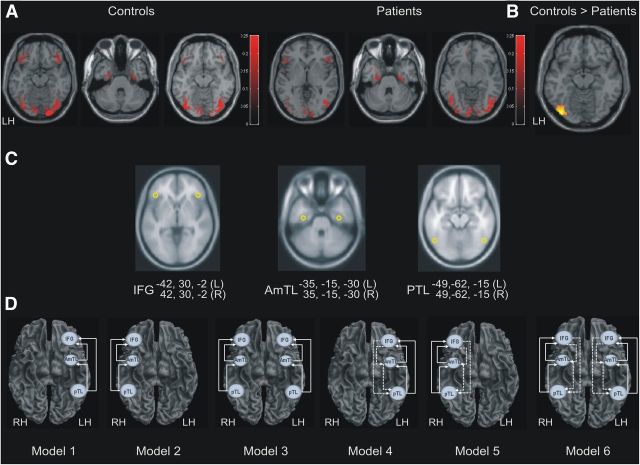
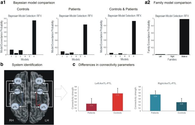
An increasing amount of evidence supports a crucial role for the anterior temporal lobe (ATL) in semantic processing. Critically, a selective disruption of the functional connectivity between left and right ATLs in patients with chronic aphasic stroke has been illustrated. The aim of the current study was to evaluate the consequences that lesions on the ATL have on the neurocognitive network supporting semantic cognition. Unlike previous work, in this magnetoencephalography study we selected a group of patients with small lesions centered on the left anteroventral temporal lobe before surgery. We then used an effective connectivity method (i.e., dynamic causal modeling) to investigate the consequences that these lesions have on the functional interactions within the network. This approach allowed us to evaluate the directionality of the causal interactions among brain regions and their associated connectivity strengths. Behaviorally, we found that semantic processing was altered when patients were compared with a strictly matched group of controls. Dynamic causal modeling for event related responses revealed that picture naming was associated with a bilateral frontotemporal network, encompassing feedforward and feedback connections. Comparison of specific network parameters between groups revealed that patients displayed selective network adjustments. Specifically, backward connectivity from anterior to posterior temporal lobe was decreased in the ipsilesional hemisphere, whereas it was enhanced in the contralesional hemisphere. These results reinforce the relevance of ATL in semantic memory, as well as its amodal organization, and highlight the role of feedback connections in enabling the integration of the semantic information.


Figures

Figure 1.
Figure 1.
Representative images of different lesion types involving the left temporobasal area for each patient are shown. Note that most of these lesions are subtle and easily missed if not guided by semiology and EEG findings. Patient 1, Axial T1 image shows a small encephalocele at the basal tip of the left temporal lobe. Patient 2,Coronal FLAIR image shows abnormal gyration pattern at the temporobasal surface. Note there is blurring of the gray-white matter encompassing entorhinal and perirhinal cortices (Insausti et al., 1998; Frankó et al., 2013) suggestive of a focal cortical dysplasia (P.2A). PET scan coregistered with MRI shows a circumscribed area of hypometabolism involving these two gyri (P.2B). Patient 3, Axial T2 image shows a small encephalocele that distorts the normal morphology of the temporal pole. Patient 4, Coronal T1 image displays asymmetrical temporal lobes with an unusual morphology of the left temporal pole that appears atrophic (P.4A). PET scan coregistered to MRI shows a subtle area of focal hypometabolism in the same localization (P.4B). Patient 5, Axial T2 image acquired along the hippocampal axis shows a small encephalocele that distorts the normal morphology of the temporal pole. Patient 6, Sagittal T2-weighted section displays a pedunculated lesion from the basal tip of the left temporal lobe. The lesion shown corresponds with a small encephalocele that is better identified with a fat saturation sequence. Patient 7, Axial FLAIR section through the temporal tip shows a small area of hyperintensity localized to the inner part of the temporal pole (P.7A). CT scan coregistered with the MRI confirmed a bone defect with the same localization (P.7B). PET scan coregister with the CT scan displays again the minor skull defect together with basal hypomebolism at the tip of the left temporal lobe (P.7C). Patient 8, Coronal T1 image displays asymmetrical temporal lobes with an unusual morphology of the left temporal pole that appears atrophic. Patient 9, Axial T1 parallel to the hippocampus plane demonstrating subtle lobulated appearance of the left temporal pole (P.9A). Fusion of the MRI and CT scans with fine axial cuts identified this abnormality as a small anterobasal temporal encephalocele (P.9B). A bone defect of the inner lamina of the skull at the medial aspect of the middle cranial fossa is evident on CT scan (P.9C). Patient 10, Coronal T2 image shows a cavernous angioma localized at the posterior portion of entorhinal/perirhinal cortex (Insausti et al., 1998; Frankó et al., 2013).
Figure 2.
Figure 2.
A, Axial views of the source localization for the grand-mean responses averaged over controls (left) and over patients (right) projected into MNI voxel space and superimposed on the template structural MRI image. B, Posterior temporo-occipital region showing significantly increased activity in controls relative to patients. C, Sources of activity, modeled as dipoles (estimated posterior moments and locations) superimposed on an MRI of a standard brain in MNI space, and their coordinates. D, Outline of the six DCM models for the effective connectivity analysis shown on axial brain schematics. Models differed in hierarchical levels (i.e., sources and extrinsic connections). Model sources could be unilateral (left or right), or bilateral. Arrows between the regions indicate the directionality of the connections feedforward or backward (dashed lines). IFG, Inferior frontal gyrus; AmTL, anteromedial temporal lobe; PTL, posterior temporal lobe; LH, left hemisphere; RH, right hemisphere.
Figure 3.
Figure 3.
a1, Bayesian model selection among the six models; and between families of models (a2). Random fixed effects (RFX) showed model expected probability and model exceedance probability. b, results indicate that Model 6 had the greatest evidence (exceedance probability = 0.931); and its subject-specific parameters (restricted to posterior probabilities of 90% or more) were selected to test for group differences (c). Red indicates differences in left hemisphere and blue indicates differences in right hemisphere.

References

    1. Agosta F, Henry RG, Migliaccio R, Neuhaus J, Miller BL, Dronkers NF, Brambati SM, Filippi M, Ogar JM, Wilson SM, Gorno-Tempini ML. Language networks in semantic dementia. Brain. 2010;133:286–299. doi: 10.1093/brain/awp233. - DOI - PMC - PubMed
    1. Antonucci SM, Beeson PM, Labiner DM, Rapcsak SZ. Lexical retrieval and semantic knowledge in patients with left inferior temporal lobe lesions. Aphasiology. 2008;22:281–304. doi: 10.1080/02687030701294491. - DOI - PMC - PubMed
    1. Bar M, Kassam KS, Ghuman AS, Boshyan J, Schmidt AM, Dale AM, Hämäläinen MS, Marinkovic K, Schacter DL, Rosen BR, Halgren E. Top-down facilitation of visual recognition. Proc Natl Acad Sci U S A. 2006;103:449–454. doi: 10.1073/pnas.0507062103. - DOI - PMC - PubMed
    1. Barense MD, Rogers TT, Bussey TJ, Saksida LM, Graham KS. Influence of conceptual knowledge on visual object discrimination: insights from semantic dementia and MTL amnesia. Cereb Cortex. 2010;20:2568–2582. doi: 10.1093/cercor/bhq004. - DOI - PubMed
    1. Bianchi AM, Marchetta E, Tana MG, Tettamanti M, Rizzo G. Frequency-based approach to the study of semantic brain networks connectivity. J Neurosci Methods. 2013;212:181–189. doi: 10.1016/j.jneumeth.2012.10.005. - DOI - PubMed

Publication types

MeSH terms