Review
. 2013 Oct;92(10):860-7.
doi: 10.1177/0022034513500306. Epub 2013 Aug 1.

Advances in the regulation of osteoclasts and osteoclast functions

Affiliations
Review

Advances in the regulation of osteoclasts and osteoclast functions

B F Boyce. J Dent Res. 2013 Oct.

Abstract

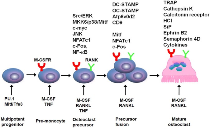
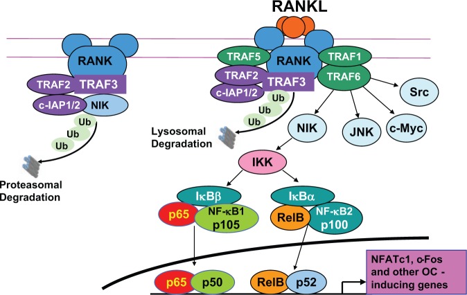
Osteoclasts are derived from mononuclear hematopoietic myeloid lineage cells, which are formed in the bone marrow and are attracted to the bloodstream by factors, including sphingsine-1 phosphate. These circulating precursors are attracted to bone surfaces undergoing resorption by chemokines and other factors expressed at these sites, where they fuse to form multinucleated bone-resorbing cells. All aspects of osteoclast formation and functions are regulated by macrophage-colony-stimulating factor (M-CSF) and receptor activator of NF-κB ligand (RANKL), cytokines essential for osteoclast formation and expressed by a variety of cell types, including osteoblast lineage cells. Since the discovery of RANKL in the mid-1990s, mouse genetic and molecular studies have revealed numerous signaling pathways activated by RANKL and M-CSF. More recent studies indicate that osteoclasts and their precursors regulate immune responses and osteoblast formation and functions by means of direct cell-cell contact through ligands and receptors, such as ephrins and Ephs, and semaphorins and plexins, and through expression of clastokines. There is also growing recognition that osteoclasts are immune cells with roles in immune responses beyond mediating the bone destruction that can accompany them. This article reviews recent advances in the understanding of the molecular mechanisms regulating osteoclast formation and functions and their interactions with other cells in normal and pathologic states.

Keywords: apoptosis; bone biology; bone remodeling/regeneration; chemokine(s); cytokine(s); osteoblast(s).


Conflict of interest statement

The author declares no potential conflicts of interest with respect to the authorship and/or publication of this article.

Figures

Figure 1.
Figure 1.
Regulation of osteoclast formation and differentiation. An early requirement for myeloid progenitor differentiation into osteoclast precursors is expression of PU.1 and Mitf, which induce expression of M-CSFR, the receptor for M-CSF. M-CSF induces expression of RANK on these cells, priming them for further differentiation in response to RANKL. TNF also induces expression of RANK, which can enhance osteoclast formation in inflammatory bone disease. In response to RANKL and TNF, expression of a number of transcription factors that regulate further differentiation of RANK-expressing cells is increased. These include NF-κB, c-Fos, and NFATc1. They induce expression of several gene-encoding proteins involved in osteoclast activation, including tartrate-resistant acid (TRAP), cathepsin-K, and the calcitonin receptor, and mediate production of H+ and Cl-, which form HCl under the ruffled borders of osteoclasts. RANKL and TNF also induce activation of c-myc, which promotes further proliferation of these cells, as well as map kinase/kinase 6 (MKK6), p38, and Mitf, along with Src and Erk, which have multiple effects to activate osteoclasts and promote their survival.
Figure 2.
Figure 2.
Functions of TRAFs in osteoclast formation and differentiation. Under basal conditions, TRAF2, c-IAP1/2, and TRAF3 form a complex on the intracellular portion of RANK. This complex polyubiquitinates NIK, which is transported to the proteasome for degradation, resulting in very low levels of NIK in unstimulated OCPs. RANKL binding to RANK on OCPs leads to recruitment of TRAFs 1, 5, and 6 and polyubiquitination of TRAF3 by TRAF2/c-IAP1/2 with subsequent lysosomal degradation of TRAF3 (our unpublished observations). This leads to the release of NIK, and signaling through it and TRAF6 results in the activation of NF-κB and subsequent increased expression of NFATc1 and c-Fos to induce further OCP differentiation. TRAF6 also mediates the activation of signaling through JNK, c-Myc, and Src to promote OCP differentiation and activation.
Figure 3.
Figure 3.
Osteoclasts actively resorbing bone. Osteoclasts with ruffled borders (black arrows) are actively resorbing bone. Note also numerous other mononuclear cells in the marrow adjacent to the osteoclasts. These include immune and stromal cells and osteoclast precursors. H&E, orange G, and phloxine.
Figure 4.
Figure 4.
Giant cell reparative granuloma of bone. Biopsy tissue from a lytic lesion in the mandible of a 14-year-old girl, showing reactive new bone (black arrow), adjacent fibrous tissue, and numerous osteoclasts (white arrow) far removed from the bone surface. H&E.
Figure 5.
Figure 5.
Giant cell tumor of bone. Numerous enormous multinucleated osteoclasts from a giant cell tumor of bone from the distal radius of a 25-year-old man, with associated mononuclear precursors and stromal cells with no bone matrix associated with them. H&E.
Figure 6.
Figure 6.
Osteoclasts associated with a sarcoma in the lung. Multinucleated osteoclasts (red arrow) are present in close association with a spindle-cell primary sarcoma in the lung of a 67-year-old woman. Lung alveolar wall (black arrow). Tartrate-resistant acid phosphatase, counterstained with hematoxylin.

References

    1. Aarthi JJ, Darendeliler MA, Pushparaj PN. (2011). Dissecting the role of the S1P/S1PR axis in health and disease. J Dent Res 90:841-854. - PubMed
    1. Asagiri M, Takayanagi H. (2007). The molecular understanding of osteoclast differentiation. Bone 40:251-264. - PubMed
    1. Barrow AD, Raynal N, Andersen TL, Slatter DA, Bihan D, Pugh N, et al. (2011). OSCAR is a collagen receptor that costimulates osteoclastogenesis in DAP12-deficient humans and mice. J Clin Invest 121:3505-3516. - PMC - PubMed
    1. Berthelot JM, Le Goff B. (2010). Rheumatoid arthritis and periodontal disease. Joint Bone Spine 77:537-541. - PubMed
    1. Boonen S, Rosenberg E, Claessens F, Vanderschueren D, Papapoulos S. (2012). Inhibition of cathepsin K for treatment of osteoporosis. Curr Osteoporos Rep 10:73-79. - PubMed

Publication types

LinkOut - more resources