Comparative Study
. 2013;42(9):20130157.
doi: 10.1259/dmfr.20130157. Epub 2013 Aug 1.

Pre-surgical treatment planning of maxillary canine impactions using panoramic vs cone beam CT imaging

Affiliations
Comparative Study

Pre-surgical treatment planning of maxillary canine impactions using panoramic vs cone beam CT imaging

A Alqerban et al. Dentomaxillofac Radiol. 2013.

Abstract

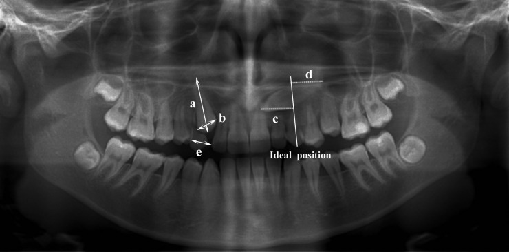
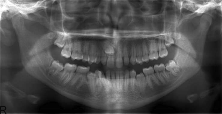
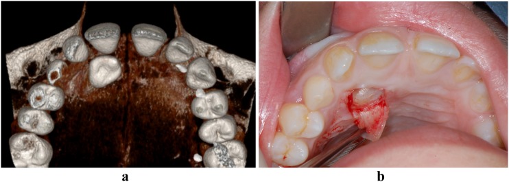
Objectives: The aim of this prospective study was to compare the impact of using two-dimensional (2D) panoramic radiographs and three-dimensional (3D) cone beam CT for the surgical treatment planning of impacted maxillary canines.

Methods: This study consisted of 32 subjects (19 females, 13 males) with a mean age of 25 years, referred for surgical intervention of 39 maxillary impacted canines. Initial 2D panoramic radiography was available, and 3D cone beam CT imaging was obtained upon clinical indication. Both 2D and 3D pre-operative radiographic diagnostic sets were subsequently analysed by six observers. Perioperative evaluations were conducted by the treating surgeon. McNemar tests, hierarchical logistic regression and linear mixed models were used to explore the differences in evaluations between imaging modalities.

Results: Significantly higher confidence levels were observed for 3D image-based treatment plans than for 2D image-based plans (p < 0.001). The evaluations of canine crown position, contact relationship and lateral incisor root resorption were significantly different between the 2D and 3D images. By contrast, pre- and perioperative evaluations were not significantly different between the two image modalities.

Conclusions: Surgical treatment planning of impacted maxillary canines was not significantly different between panoramic and cone beam CT images.

Keywords: cone beam CT; cuspid; impacted; panoramic; teeth.


Figures

Figure 1
Figure 1
Panoramic image of 15 year old female patient with bilateral impacted canine illustrating the reference lines of ideal canine position as well as the linear measurements as follows: (a) the canine total length from the canine cusp tip to the apex, (b) the canine crown width, (c) the distance from the canine cusp tip perpendicular to the axis of the ideal position, (d) the distance from the canine apex perpendicular to the axis of the ideal position and (e) the mesiodistal space from the distal surface of the lateral incisor to the mesial surface of the first premolar
Figure 2
Figure 2
Two-dimensional panoramic radiograph of a 15 year old female with an impacted maxillary right canine. The root contours of the central and lateral incisors overlap with that of the canine. The canine crown is magnified indicating that it is palatally impacted with the exact location for surgical intervention is very difficult to assess
Figure 3
Figure 3
(a) Three-dimensional cone beam CT image from the 3D Accuitomo-XYZ Slice View Tomograph® system (J. Morita, Kyoto, Japan) showing the exact location of the right maxillary canine. (b) Occlusal intraoral photograph showing the crown of the impacted upper right maxillary canine during surgery

References

    1. Bishara SE. Impacted maxillary canines: a review. Am J Orthod Dentofacial Orthop 1992; 101: 159–171 - PubMed
    1. Ericson S, Kurol J. Radiographic examination of ectopically erupting maxillary canines. Am J Orthod Dentofacial Orthop 1987; 91: 483–492 - PubMed
    1. Peck S, Peck L, Kataja M. The palatally displaced canine as a dental anomaly of genetic origin. Angle Orthod 1994; 64: 249–256 10.1043/0003-3219(1994)064<0249:WNID>2.0.CO;2 - DOI - PubMed
    1. Kim Y, Hyun HK, Jang KT. The position of maxillary canine impactions and the influenced factors to adjacent root resorption in the Korean population. Eur J Orthod 2012; 34: 302–306 10.1093/ejo/cjr002 - DOI - PubMed
    1. Alqerban A, Jacobs R, Lambrechts P, Loozen G, Willems G. Root resorption of the maxillary lateral incisor caused by impacted canine: a literature review. Clin Oral Investig 2009; 13: 247–255 10.1007/s00784-009-0262-8 - DOI - PubMed

Publication types

MeSH terms