

Carotid CTA: radiation exposure and image quality with the use of attenuation-based, automated kilovolt selection
- PMID: 23907241
- PMCID: PMC7965763
- DOI: 10.3174/ajnr.A3659
Carotid CTA: radiation exposure and image quality with the use of attenuation-based, automated kilovolt selection
Abstract
Background and purpose: CTA is considered the imaging modality of choice in evaluating the supraaortic vessels in many institutions, but radiation exposure remains a matter of concern. The objective of the study was to evaluate a fully automated, attenuation-based kilovolt selection algorithm in carotid CTA in respect to radiation dose and image quality compared with a standard 120-kV protocol.
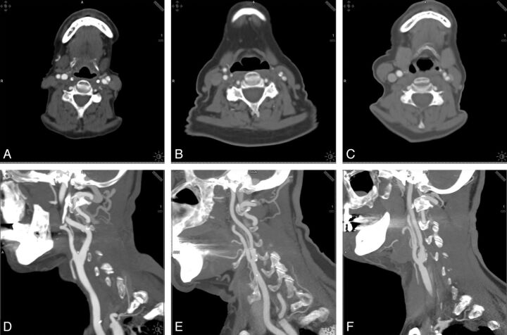
Materials and methods: Ninety-eight patients were included: 53 examinations (patient age, 66 ± 12 years) were performed by use of automated adaption of tube potential (80-140 kV) on the basis of the attenuation profile of the scout scan (study group), and 45 examinations (patient age, 67 ± 11 years) were performed by use of a standard 120-kV protocol (control group). CT dose index volume and dose-length product were recorded from the examination protocol. Image quality was assessed by ROI measurements and calculations of SNR and contrast-to-noise ratio. Subjective image quality was evaluated by 2 observers with the use of a 4-point scale (3, excellent; 0, not diagnostic).
Results: Subjective image quality was rated as "excellent" or "good" in all examinations (study group, 2.8; control group, 2.8). The algorithm automatically selected 100 kV in 47% and 80 kV in 34%; 120 kV was retained in 19%. An elevation to 140 kV did not occur. Compared with the control group, overall CT dose index volume reduction was 33.7%; overall dose-length product reduction was 31.5%. In the low-kilovolt scans, image noise and mean attenuation of ROIs inside the carotid arteries were significantly higher than in 120-kV scans, resulting in a constant or increased (80-kV group) contrast-to-noise ratio.
Conclusions: The attenuation-based, kilovolt selection algorithm enables a dose reduction of >30% in carotid artery CTA while maintaining contrast-to-noise ratio and subjective image quality at adequate levels.
Figures
References
-
- Brott TG, Halperin JL, Abbara S, et al. 2011 ASA/ACCF/AHA/AANN/AANS/ACR/ASNR/CNS/SAIP/SCAI/SIR/SNIS/SVM/SVS Guideline on the management of patients with extracranial carotid and vertebral artery disease: a report of the American College of Cardiology Foundation/American Heart Association Task Force on Practice Guidelines, and the American Stroke Association, American Association of Neuroscience Nurses, American Association of Neurological Surgeons, American College of Radiology, American Society of Neuroradiology, Congress of Neurological Surgeons, Society of Atherosclerosis Imaging and Prevention, Society for Cardiovascular Angiography and Interventions, Society of Interventional Radiology, Society of NeuroInterventional Surgery, Society for Vascular Medicine, and Society for Vascular Surgery, developed in collaboration with the American Academy of Neurology and Society of Cardiovascular Computed Tomography. J Am Coll Cardiol 2011;57:e16–94 - PubMed
-
- Kuefner MA, Grudzenski S, Schwab SA, et al. DNA double-strand breaks and their repair in blood lymphocytes of patients undergoing angiographic procedures. Invest Radiol 2009;44:440–46 - PubMed
-
- Brenner DJ, Hall EJ. Computed tomography: an increasing source of radiation exposure. N Engl J Med 2007;357:2277–84 - PubMed
Publication types
MeSH terms
LinkOut - more resources
Full Text Sources
Other Literature Sources
Medical