

Hyperattenuated intracerebral lesions after mechanical recanalization in acute stroke
- PMID: 23907245
- PMCID: PMC7965757
- DOI: 10.3174/ajnr.A3656
Hyperattenuated intracerebral lesions after mechanical recanalization in acute stroke
Abstract
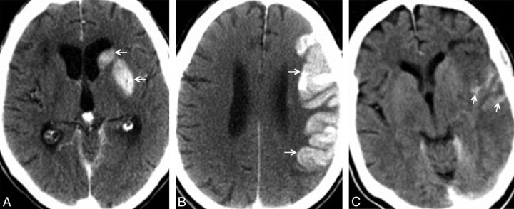
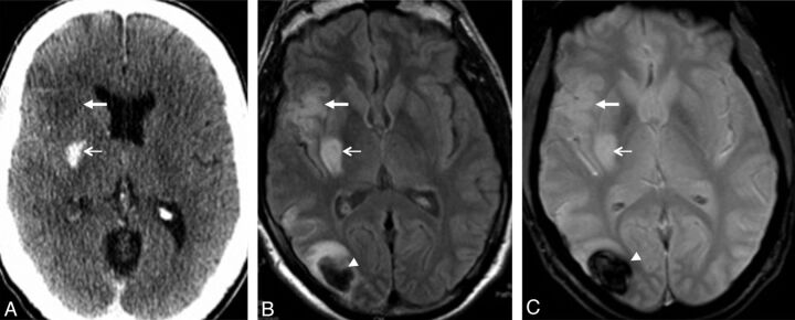
Background and purpose: Following mechanical recanalization of an acute intracranial vessel occlusion, hyperattenuated lesions are frequently found on postinterventional cranial CT. They represent either blood or-more frequently-enhancement of contrast agent. Here, we aimed to evaluate the prognostic value of these hyperattenuated intracerebral lesions.
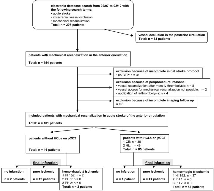
Materials and methods: One hundred one consecutive patients with acute stroke in the anterior circulation who underwent mechanical recanalization were included. Risk factors for hyperattenuated intracerebral lesions were assessed, and lesion volume was compared with the volume of final infarction. Clinical outcome and relative risk of secondary hemorrhage were determined in patients with and without any hyperattenuated lesions and compared.
Results: The frequency of hyperattenuated lesions was 84.2%. Risk factors for hyperattenuated lesions were female sex, higher NIHSS score on admission, and higher amount of contrast agent applied. On follow-up, 3 patients showed no infarction; 53 patients, an ischemic infarction; and 45 patients, a hemorrhagic infarction. In all except 1 case, final volume of infarction (median = 92.4 mL) exceeded the volume of hyperattenuated intracerebral lesions (median = 5.6 mL). Patients with hyperattenuated lesions were at a 4 times higher relative risk for hemorrhagic transformation but had no significantly worse clinical outcome.
Conclusions: Our data show that the extent of postinterventional hyperattenuated intracerebral lesions underestimates the volume of final infarction. Although hyperattenuated lesions indicate a higher risk of secondary hemorrhagic transformation, their presence seems not to be of any prognostic value regarding clinical outcome.
Figures
Comment in
-
Hyperattenuated intracerebral lesions after mechanical recanalization in acute stroke: contrast and compare.AJNR Am J Neuroradiol. 2014 Feb;35(2):352-3. doi: 10.3174/ajnr.A3824. Epub 2013 Nov 28. AJNR Am J Neuroradiol. 2014. PMID: 24287093 Free PMC article. No abstract available.
References
-
- Komiyama M, Nishijima Y, Nishio A, et al. Extravasation of contrast medium from the lenticulostriate artery following local intracarotid fibrinolysis. Surg Neurol 1993;39:315–19 - PubMed
-
- Nakano S, Iseda T, Kawano H, et al. Parenchymal hyperdensity on computed tomography after intra-arterial reperfusion therapy for acute middle cerebral artery occlusion: incidence and clinical significance. Stroke 2001;32:2042–48 - PubMed
-
- Yoon W, Seo JJ, Kim JK, et al. Contrast enhancement and contrast extravasation on computed tomography after intra-arterial thrombolysis in patients with acute ischemic stroke. Stroke 2004;35:876–81 - PubMed
MeSH terms
LinkOut - more resources
Full Text Sources
Other Literature Sources
Medical