. 2013 Aug 23;341(6148):893-6.
doi: 10.1126/science.1241530. Epub 2013 Aug 1.

Titration of four replication factors is essential for the Xenopus laevis midblastula transition

Affiliations

Titration of four replication factors is essential for the Xenopus laevis midblastula transition

Clara Collart et al. Science. .

Abstract

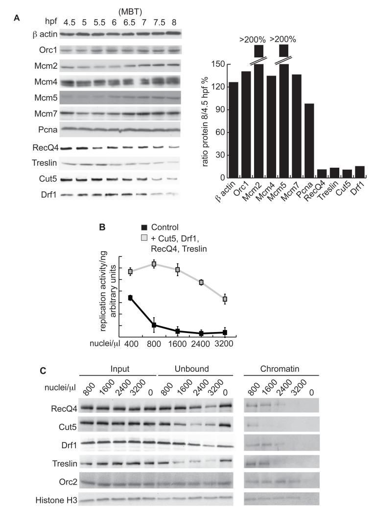
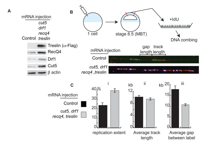
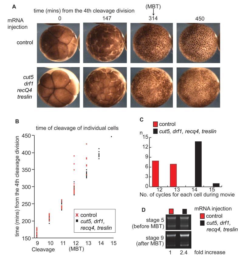
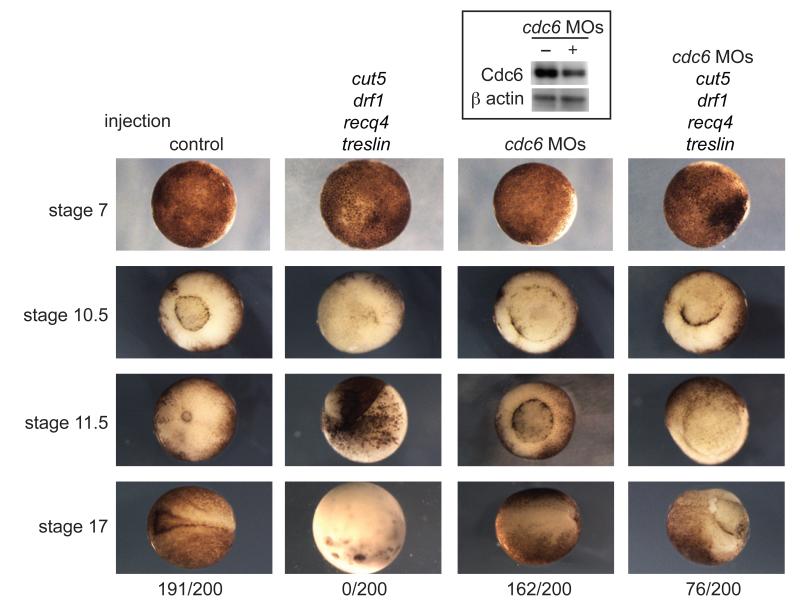
The rapid, reductive early divisions of many metazoan embryos are followed by the midblastula transition (MBT), during which the cell cycle elongates and zygotic transcription begins. It has been proposed that the increasing nuclear to cytoplasmic (N/C) ratio is critical for controlling the events of the MBT. We show that four DNA replication factors--Cut5, RecQ4, Treslin, and Drf1--are limiting for replication initiation at increasing N/C ratios in vitro and in vivo in Xenopus laevis. The levels of these factors regulate multiple events of the MBT, including the slowing of the cell cycle, the onset of zygotic transcription, and the developmental activation of the kinase Chk1. This work provides a mechanism for how the N/C ratio controls the MBT and shows that the regulation of replication initiation is fundamental for normal embryogenesis.


Figures

Figure 1
Figure 1. Initiation factors are limiting for replication in vitro in Xenopus laevis
A. (Left) Western blot of DNA replication factors from Xenopus embryos at the indicated hours post fertilization (hpf) (. The MBT occurs at 6 hours 30 mins (6.5). (Right) Quantification of western blots using Image J. Protein levels are represented as the ratio between 8/4.5 hpf. Pcna is required for DNA replication elongation and did not change in abundance during early embryogenesis. B. Replication of Xenopus sperm chromatin nuclei in IVT diluted egg extract. Sperm chromatin was added at the indicated concentrations either supplemented with rabbit reticulocyte lysate + empty vector (control) or + vectors encoding cut5, drf1, recq4 and treslin (grey). The reactions were quantified relative to input DNA to represent replication activity per ng of sperm DNA. n=3, error bars show standard deviation. C. Western blot of chromatin binding of replication factors at increasing sperm nuclei to extract ratios. Xenopus sperm nuclei of the indicated ratios were replicated for 15 mins in undiluted egg extract. Input and chromatin unbound fractions were equally loaded by volume, while the chromatin fractions were normalized to the same DNA concentration.
Figure 2
Figure 2. Cut5, Drf1, RecQ4 and Treslin are limiting for replication in vivo in Xenopus laevis
A. Western blot from stage 9 (post MBT) embryos either injected at the 1 cell stage with water (control) or with 300 pg mRNA each of treslin, cut5, recq4, and drf1. B. (Top) sSchematic representation of experimental design. Xenopus embryos were injected at the one cell stage as in A in the animal pole. At 6.5 hpf (stage 8.5) animal caps were cut and their cells dissociated. The cells were given a pulse with IdU and incorporation was visualized by anti IdU immunofluorescence after stretching DNA onto slides (bottom, DNA-red, IdU-green). C. Analysis from B of (i) replication extent (%), (ii) average IdU track length (kb) and (iii) average gap between label (kb). n = 96 and 93 fibers for control and overexpressing embryos, respectively.
Figure 3
Figure 3. Overexpression of Cut5, Drf1, RecQ4 and Treslin causes extra rapid cell divisions after the MBT
A. Images taken from a time-lapse movie (Movie S1) of embryos injected at the 1 cell stage as in Fig.2A. Cleavage 4 at the 16-cell stage was set to time zero. B. 15 individual cells from the embryos in Movie S1 were followed through early divisions until 450 minutes after cleavage 4. Each time point corresponds to the cleavage of an individual cell. Cleavages 1-8 are excluded for simplicity. C. Representation of the number of cell cycles each cell in B undergoes until 450 minutes after cleavage 4. D. Embryos were injected as in A and half the DNA content of a single embryo at stages 5 and 9 was loaded onto an agarose gel and stained with ethidium bromide. This gel is representative of triplicate repeats. Below - DNA amount at stage 9 was quantified using ImageJ and fold increase relative to control is represented.
Figure 4
Figure 4. Increasing inter-origin distances at the MBT is critical for gastrulation and neurulation
Images of embryos injected with water (control - first column), treslin, cut5, recq4, and drf1 mRNA (second column), cdc6 morpholinos (MOs – third column) and embryos injected with a mixture of the cdc6 morpholinos and mRNA for treslin, cut5, recq4, and drf1 (fourth column). Stage 7 = pre-MBT, 10.5 = gastrulation, 11.5 = mid-gastrulation, 17 = neurula. The number of embryos out of 200 surviving at stage 17 is indicated at the bottom. (Top) Western blot of knockdown of Cdc6 after morpholino injection.

Comment in

Similar articles

Cited by

References

    1. Tadros W, Lipshitz HD. Development. 2009 Sep;136:3033. - PubMed
    1. Shimuta K, et al. EMBO J. 2002 Jul 15;21:3694. - PMC - PubMed
    1. Sibon OC, Stevenson VA, Theurkauf WE. Nature. 1997 Jul 3;388:93. - PubMed
    1. Newport J, Kirschner M. Cell. 1982 Oct;30:675. - PubMed
    1. Newport J, Kirschner M. Cell. 1982 Oct;30:687. - PubMed

Publication types