. 2013 Oct;4(5):569-79.
doi: 10.1007/s13244-013-0271-7. Epub 2013 Aug 2.

Skeletal involvement in Langerhans cell histiocytosis

Affiliations

Skeletal involvement in Langerhans cell histiocytosis

Suonita Khung et al. Insights Imaging. 2013 Oct.

Abstract

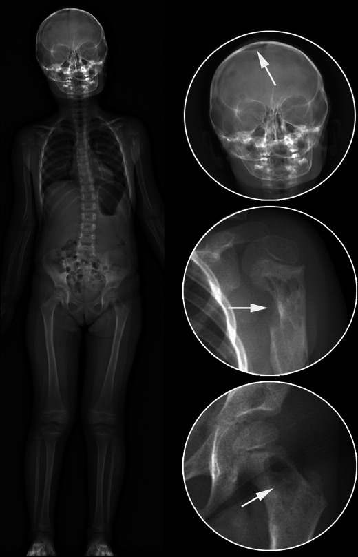
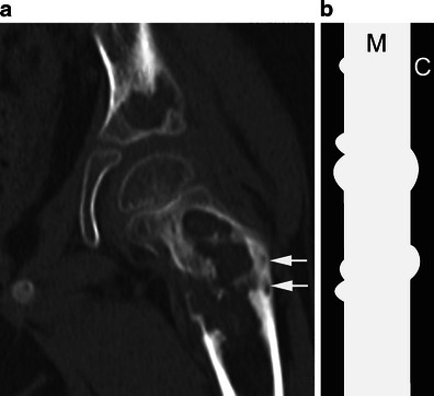
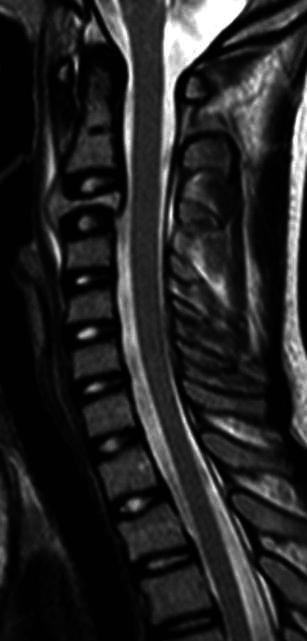
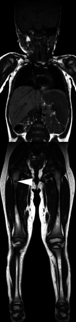
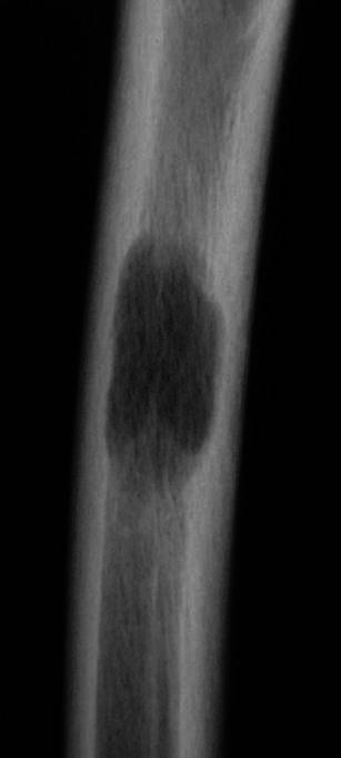
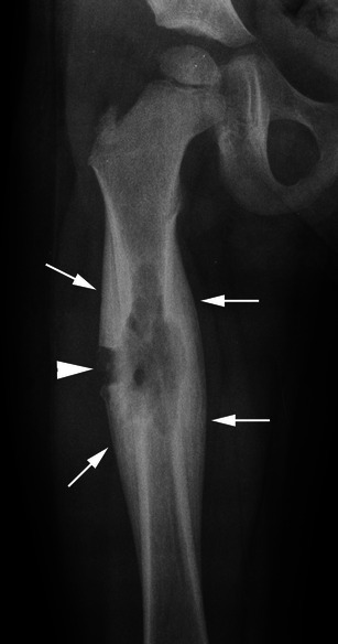
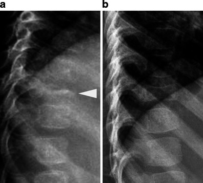
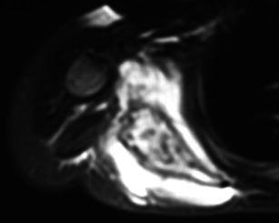
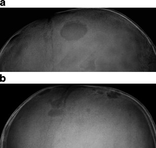
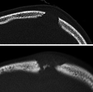
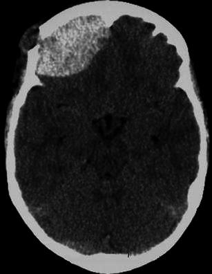
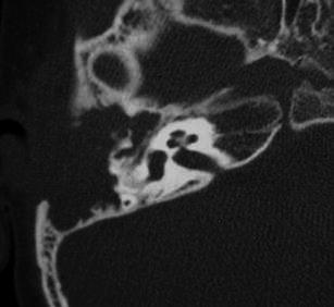
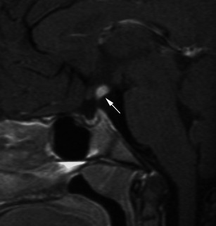
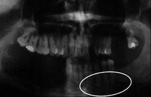
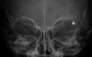
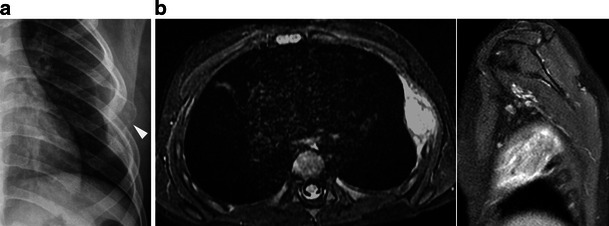
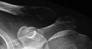
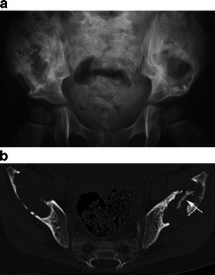
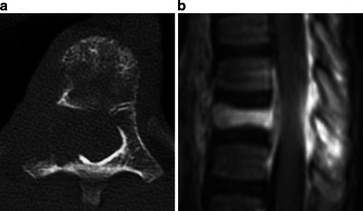
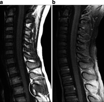
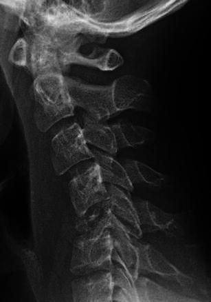
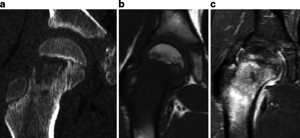
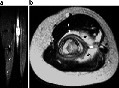
Langerhans cell histiocytosis (LCH) represents a disorder characterised by an abnormal accumulation of histiocytes in miscellaneous tissues. The bone is commonly affected, especially the flat bones, the spine and the long bones. Some lesions in children such as a "vertebra plana" or a solitary lytic lesion of the skull may be suggestive for LCH, whereas others can be confused with a malignant tumour or osteomyelitis. This pictorial essay presents the main usual and unusual skeletal manifestations observed in LCH.

Teaching points: • Osseous involvement in children with LCH is very similar to that seen in multiple myeloma. • A solitary lytic lesion of the cranial vault is a typical radiographic finding of LCH. • A vertebra plana appearance in the spine is another typical radiographic finding. • Extensive signal intensity changes within bone marrow on MRI are a helpful sign for the diagnosis. • In long bones, endosteal scalloping may be responsible for a "budding appearance".


Figures

Fig. 1
Fig. 1
Low-dose biplanar skeletal survey (anteroposterior view) reveals bone lesions (arrows) of the calvarium, the proximal left humerus and the proximal left femur
Fig. 2
Fig. 2
a Coronal CT image of the left hip shows large, well-defined, lytic, femoral and acetabular lesions. The femoral lesion, at risk of impending fracture, was immobilised thereafter. Note the “budding appearance” of bone destruction (arrows) related to endosteal scalloping. b The diagram depicts this “budding appearance” in the diaphysis of a long bone. M medullary canal, C cortex
Fig. 3
Fig. 3
Sagittal T2-weighted MR image of the cervical spine demonstrates a lesion of C3 associated with severe vertebral collapse and prevertebral and epidural soft tissue extension. There is no compression of the spinal cord
Fig. 4
Fig. 4
Coronal whole-body T1-weighted MR image reveals a right femoral bone lesion (arrowhead) and splenomegaly. Courtesy of Pr Guy Sebag and Dr Marianne Alison, Paris, France
Fig. 5
Fig. 5
Anteroposterior radiograph of a femur shows an ill-defined lytic medullary lesion
Fig. 6
Fig. 6
Anteroposterior radiograph of the right femur reveals ill-defined trabecular osteolysis associated with periosteal buttressing (arrows), indicating a relatively slow-growing lesion. A bone biopsy (arrowhead) confirmed the diagnosis
Fig. 7
Fig. 7
a Lateral radiographs of the spine show a complete vertebral collapse of T10 (vertebra plana appearance) (arrowhead) with preservation of the adjacent disc spaces. b Partial height restitution is seen 1 year later
Fig. 8
Fig. 8
Axial T2-weighted MR image reveals extensive signal intensity changes within the bone marrow of the right scapula and the adjacent soft tissues. The lesion is markedly expansile
Fig. 9
Fig. 9
Lateral skull radiographs demonstrate one (a) or several (b) well-defined lytic lesions of the skull in the frontal and parietal bones
Fig. 10
Fig. 10
Axial CT scans of two calvarial lesions show bevelled margins due to unequal destruction of the inner and outer tables of the skull and a “button” sequestrum (i.e. a fragment of residual bone within a lytic lesion)
Fig. 11
Fig. 11
Axial unenhanced CT scan exhibits an extra-dural hematoma related to the bleeding of a right frontal bone lesion
Fig. 12
Fig. 12
Axial CT scan of the right mastoid in a child presenting with otorrhea demonstrates an important destruction of the mastoid part of the right temporal bone and the middle ear, with partial lysis of the ossicular chain. Complete reossification of the temporal bone was seen on CT 2 years later
Fig. 13
Fig. 13
Sagittal fat-suppressed enhanced T1-weighted MR image shows nodular enhancement of the infundibular pituitary stalk (arrow)
Fig. 14
Fig. 14
Dental panoramic radiograph in a young adult shows a left mandibular lesion. The destruction of the alveolar bone gives the appearance of “floating teeth”
Fig. 15
Fig. 15
Anteroposterior radiograph of the skull shows osteolysis of the lateral part of the left orbit (asterisk)
Fig. 16
Fig. 16
a Chest radiograph reveals an expansile and lytic lesion of the lateral portion of a left rib (arrowhead). 16b Axial fat-suppressed T2-weighted MR image and sagittal fat-suppressed gadolinium-enhanced T1-weighted MR image show extensive bone marrow changes with soft tissue extension creating an extrapleural mass
Fig. 17
Fig. 17
Anteroposterior radiograph of the left clavicle in a young adult shows an ill-defined, slightly expansile, lytic lesion of the lateral part of the bone
Fig. 18
Fig. 18
a Anteroposterior radiograph of the pelvis reveals bilateral lytic lesions of the iliac wings. On the left, well-defined sclerotic margins are seen around the lesion that extends to the supra-acetabular region. b On axial CT scan, note the presence of a fragment of intact bone within the left bone lesion (arrow) with reactive sclerosis
Fig. 19
Fig. 19
a Axial CT scan demonstrates an ill-defined osteolysis of a thoracic vertebra involving the vertebral body but also the right pedicle and the transverse process. b Sagittal fat-suppressed gadolinium-enhanced T1-weighted MR image shows the degree of epidural extension. The collapsed vertebral body is evident
Fig. 20
Fig. 20
Sagittal unenhanced T1- (a) and fat-suppressed gadolinium-enhanced T1-weighted (b) MR images reveal an involvement limited to the posterior elements
Fig. 21
Fig. 21
Lateral radiograph shows marked destruction of C5 body responsible for cervical kyphosis
Fig. 22
Fig. 22
a Coronal CT scan (22a) demonstrates an osteolytic lesion in the metaphysis of the right femur, without involvement of the epiphysis. Note the persistence of tiny bone fragments within the lesion. b Coronal unenhanced T1- and (c) fat-suppressed gadolinium-enhanced T1-weighted MR images show non-specific but extensive signal intensity changes within the bone marrow and the adjacent soft tissues as well as reactive synovitis
Fig. 23
Fig. 23
a Coronal fat-suppressed T2-weighted MR image reveals focal bone marrow replacement within the femoral diaphysis with a “budding” appearance due to endosteal scalloping and extensive oedema within the bone marrow and the adjacent soft tissues. b Axial T2-weighted MR image in another patient shows circumferential periosteal reaction (arrows) and extensive bone marrow oedema within the bone marrow and the adjacent soft tissues (asterisks)

References

    1. Windebank K, Nanduri V. Langerhans cell histiocytosis. Arch Dis Child. 2009;94:904–908. doi: 10.1136/adc.2007.125872. - DOI - PubMed
    1. Guyot-Goubin A, Donadieu J, Barkaoui M, Bellec S, Thomas C, Clavel J. Descriptive epidemiology of childhood Langerhans cell histiocytosis in France, 2000–2004. Pediatr Blood Cancer. 2008;51:71–75. doi: 10.1002/pbc.21498. - DOI - PubMed
    1. Stull MA, Kransdorf MJ, Devaney KO. Langerhans cell histiocytosis of bone. Radiographics. 1992;12:801–823. doi: 10.1148/radiographics.12.4.1636041. - DOI - PubMed
    1. Weitzman S, Egeler RM. Langerhans cell histiocytosis: update for the pediatrician. Current Opin Pediatr. 2008;20:23–29. doi: 10.1097/MOP.0b013e3282f45ba4. - DOI - PubMed
    1. Leonidas JC, Guelfguat M, Valderrama E. Langerhans’ cell histiocytosis (203) Lancet 361:1293–1295 - PubMed

LinkOut - more resources