. 2013 Sep;123(9):3902-13.
doi: 10.1172/JCI69485. Epub 2013 Aug 1.

Prolactin promotes cartilage survival and attenuates inflammation in inflammatory arthritis

Affiliations

Prolactin promotes cartilage survival and attenuates inflammation in inflammatory arthritis

Norma Adán et al. J Clin Invest. 2013 Sep.

Abstract

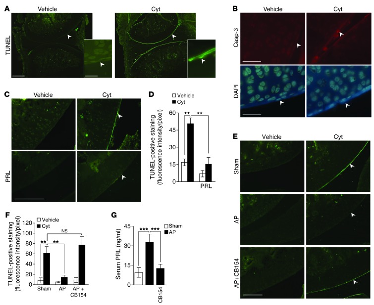
Chondrocytes are the only cells in cartilage, and their death by apoptosis contributes to cartilage loss in inflammatory joint diseases, such as rheumatoid arthritis (RA). A putative therapeutic intervention for RA is the inhibition of apoptosis-mediated cartilage degradation. The hormone prolactin (PRL) frequently increases in the circulation of patients with RA, but the role of hyperprolactinemia in disease activity is unclear. Here, we demonstrate that PRL inhibits the apoptosis of cultured chondrocytes in response to a mixture of proinflammatory cytokines (TNF-α, IL-1β, and IFN-γ) by preventing the induction of p53 and decreasing the BAX/BCL-2 ratio through a NO-independent, JAK2/STAT3-dependent pathway. Local treatment with PRL or increasing PRL circulating levels also prevented chondrocyte apoptosis evoked by injecting cytokines into the knee joints of rats, whereas the proapoptotic effect of cytokines was enhanced in PRL receptor-null (Prlr(-/-)) mice. Moreover, eliciting hyperprolactinemia in rats before or after inducing the adjuvant model of inflammatory arthritis reduced chondrocyte apoptosis, proinflammatory cytokine expression, pannus formation, bone erosion, joint swelling, and pain. These results reveal the protective effect of PRL against inflammation-induced chondrocyte apoptosis and the therapeutic potential of hyperprolactinemia to reduce permanent joint damage and inflammation in RA.


Figures

Figure 1
Figure 1. PRL inhibits Cyt-induced apoptosis of chondrocytes in culture.
(A) Primary cultures of rat chondrocytes were challenged with Cyt, combined or not with different concentrations of PRL, and apoptosis was evaluated at 24 hours by ELISA (n = 3–6). (B) Representative Western blot of procaspase-3 and active caspase-3 (Procasp-3 and Casp-3, respectively) in lysates of chondrocytes incubated or not with Cyt in the absence or presence of PRL for 6 hours. The graph shows the quantification of active caspase-3 by densitometry after normalization to procaspase-3 (n = 3). (C) qRT-PCR–based quantification of p53 mRNA levels (n = 3) in chondrocytes incubated or not with Cyt in the absence or presence of PRL for 24 hours. (D) Representative Western blot of BAX and BCL-2 in chondrocytes incubated or not with Cyt in the absence or presence of PRL for 4 hours. The graph shows the quantification of BAX/BCL-2 by densitometry (n = 3). Values are mean ± SEM. *P < 0.05, **P < 0.01, ***P < 0.001.
Figure 2
Figure 2. PRL inhibits Cyt-induced chondrocyte apoptosis by a NO-independent, JAK2/STAT3–dependent pathway.
(A) Apoptosis evaluated by ELISA in chondrocytes incubated with Cyt in the presence or absence of the NO inhibitor l-NAME for 24 hours (n = 3–6). (B) Western blot analysis of iNOS (n = 3) and (C) NO2 and NO3 concentrations (n = 7) after incubating or not incubating chondrocytes with Cyt in the absence or presence of PRL for 6 and 24 hours, respectively. (D) Representative Western blot of phosphorylated JAK2 (pJAK2) in chondrocytes incubated with the various treatments for 30 minutes (n = 3). (E) Representative immunostaining for total STAT3 and DAPI in cultured chondrocytes treated with or without (control) PRL (2.3 μg/ml), Cyt, or PRL and Cyt (PRL + Cyt) for 1 hour (n = 3). Scale bar: 25 μm. (F) Apoptosis evaluated by ELISA in chondrocytes incubated in the absence or presence of 100 nM STAT3 inhibitor S31-201 for 24 hours (n = 3–4). Bars represent mean ± SEM. *P < 0.05, ***P < 0.001.
Figure 3
Figure 3. PRL inhibits the apoptosis of chondrocytes induced by the intra-articular injection of Cyt.
(A and B) Apoptosis was assessed in rat knees injected with vehicle or Cyt by TUNEL and active caspase-3 staining. The bottom right images in A show enlarged views of knee cartilage. Scale bar: 500 μm (top); 25 μm (bottom). (B) Nucleic acids in chondrocytes were stained by DAPI. Scale bar: 50 μm. (C and D) TUNEL-positive staining and quantification in outer border cartilage of rat knees coinjected with Cyt and PRL (n = 4). Scale bar: 100 μm. (E and F) TUNEL-positive staining and quantification in outer border cartilages of rat knees from nontransplanted (sham) and AP-transplanted rats exposed to vehicle or Cyt, in the presence or absence of dopamine D2 receptor antagonist, CB154 (n = 4–6). (AC and E) White arrowheads indicate cartilage outer border. Scale bar: 250 μm. (G) Serum PRL levels in sham or AP-transplanted rats treated or not with CB154 (n = 5–10). Bars represent mean ± SEM. **P < 0.01, ***P < 0.001.
Figure 4
Figure 4. Cyt-induced chondrocyte apoptosis is enhanced in Prlr–/– mice.
Apoptosis was assessed by TUNEL staining in knees of Prlr–/– and Prlr+/+ mice intra-articularly injected or not with Cyt. (A and B) Both injected and (C and D) noninjected knee joints, i.e., ipsilateral and contralateral to the injection site, respectively, were analyzed. White arrowheads indicate cartilage outer border. Scale bar: 250 μm. Bars represent mean ± SEM. (n = 3–5). *P < 0.05, **P < 0.01, ***P < 0.001.
Figure 5
Figure 5. PRL and Hal prevent chondrocyte apoptosis in adjuvant-induced arthritis.
(A) Experimental design diagram: osmotic minipumps delivering PRL or subcutaneous tablets releasing Hal were placed 3 days before injecting the rats with CFA, and the experiment ended on day 21 after CFA. (B) Serum PRL levels on days 0 and 21 after CFA in PRL-treated rats (n = 3–8) and on days 0, 14, and 21 after CFA in Hal-treated rats (n = 4–8). (C) TUNEL and active caspase-3 staining of articular cartilage of knees from rats treated or not with PRL or Hal under control and CFA-injected conditions on day 21 after CFA. Scale bar: 100 μm. The graph shows the quantification of TUNEL-positive cells in articular cartilage (n = 4–8). (D) qRT-PCR–based quantification of Casp3, Bax, and p53 mRNA levels in ankle joints from PRL- and Hal-treated rats on day 21 after CFA (n = 5–14). Bars represent mean ± SEM. *P < 0.05, **P < 0.01, ***P < 0.001.
Figure 6
Figure 6. PRL reduces chondrocyte apoptosis in already arthritic rats.
(A) Experimental design diagram: osmotic minipumps delivering PRL were placed 15 days after the injection of CFA in rats, and the experiment ended on day 21 after CFA. (B) Serum PRL levels on day 21 after CFA in PRL-treated and nontreated rats (n = 4–8). (C) TUNEL and active caspase-3 staining of articular cartilage of knees from rats treated or not with PRL under control and CFA-injected conditions on day 21 after CFA. Scale bar: 100 μm. The graph shows the quantification of TUNEL-positive cells in articular cartilage (n = 5–8). (D) qRT-PCR–based quantification of Casp3, Bax, and p53 mRNA levels in ankle joints from PRL-treated and nontreated rats on day 21 after CFA (n = 3–8). Bars represent mean ± SEM. *P < 0.05, **P < 0.01, ***P < 0.001.
Figure 7
Figure 7. PRL and Hal prevent joint inflammation in adjuvant-induced arthritis.
(A) Experimental design diagram: osmotic minipumps delivering PRL or subcutaneous tablets releasing Hal were implanted 3 days before the injection of CFA in rats. (B) Representative photographs of hind paws from groups injected or not with CFA. (C and F) Time course of ankle circumference in groups infused with PRL (n = 10) or treated with Hal (n = 16) under control and CFA-injected conditions. (C) Days 15, 18, and 21, P < 0.001, CFA vs. control. Days 18 and 21, P < 0.001, PRL vs. PRL plus CFA. (F) Days 15 and 18, P < 0.001, CFA vs. control. Days 12, 15, 18, and 21, P < 0.001, CFA vs. Hal plus CFA. (D and G) Nociceptive threshold in groups infused with PRL (n = 5–9) or treated with Hal (n = 5–9). (E and H) qRT-PCR–based quantification of Infg, Il6, iNos, Il1b, and Tnfa mRNA levels in ankle joints from rats treated with PRL (n = 3–10) or with Hal (n = 3–10) under control and CFA-injected conditions on day 21 after CFA. Bars are mean ± SEM. *P < 0.05, **P < 0.01, ***P < 0.001.
Figure 8
Figure 8. PRL reduces pannus formation and bone erosion in adjuvant-induced arthritis.
Histological evaluation of (A) pannus formation and (B) bone erosion in sections of knee joints stained by hematoxylin and eosin from nonimplanted rats (control) or rats implanted with osmotic minipumps delivering PRL or with subcutaneous tablets releasing Hal 3 days before injecting or not injecting CFA; the histological evaluation was carried out on day 21 after CFA (n = 3–8). Pannus-associated regions in each group are indicated (arrows). c, cartilage; t, bone trabeculae; mc, bone marrow cavity. Scale bar: 100 μm. Graphs show histological scores for (A) pannus formation (synovial membrane hyperplasia and infiltration of leukocytes) and (B) bone erosion (thinning and destruction of bone trabeculae). Values are mean ± SEM. **P < 0.01.
Figure 9
Figure 9. PRL reduces joint inflammation in already arthritic rats.
(A) Experimental design diagram: osmotic minipumps delivering PRL were placed 15 days after the injection of CFA in rats, and the experiment ended on day 21 after CFA. (B) Time course of ankle circumference (n = 10–15) (days 18 and 21, P < 0.001, CFA vs. control), (C) evaluation of ankle joint nociceptive threshold (n = 5–8), and (D) qRT-PCR–based quantification of Ifng, Il6, iNos, Il1b, and Tnfa mRNA levels (n = 5–8) in ankle joints under control and CFA-injected conditions on day 21 after CFA. Bars are mean ± SEM. *P < 0.05, **P < 0.01, ***P < 0.001.

References

    1. McInnes IB, Schett G. Cytokines in the pathogenesis of rheumatoid arthritis. Nat Rev Immunol. 2007;7(6):429–442. doi: 10.1038/nri2094. - DOI - PubMed
    1. Schuerwegh AJ, et al. Influence of pro-inflammatory (IL-1 alpha, IL-6, TNF-alpha, IFN-gamma) and anti-inflammatory (IL-4) cytokines on chondrocyte function. Osteoarthritis Cartilage. 2003;11(9):681–687. doi: 10.1016/S1063-4584(03)00156-0. - DOI - PubMed
    1. Goggs R, et al. Apoptosis and the loss of chondrocyte survival signals contribute to articular cartilage degradation in osteoarthritis. Vet J. 2003;166(2):140–158. doi: 10.1016/S1090-0233(02)00331-3. - DOI - PubMed
    1. Christodoulou C, Choy EH. Joint inflammation and cytokine inhibition in rheumatoid arthritis. Clin Exp Med. 2006;6(1):13–19. doi: 10.1007/s10238-006-0088-5. - DOI - PubMed
    1. Keffer J, et al. Transgenic mice expressing human tumour necrosis factor: a predictive genetic model of arthritis. EMBO J. 1991;10(13):4025–4031. - PMC - PubMed

Publication types

MeSH terms