. 2013 Aug 2;341(6145):549-53.
doi: 10.1126/science.1238327.

Functional lysine modification by an intrinsically reactive primary glycolytic metabolite

Affiliations

Functional lysine modification by an intrinsically reactive primary glycolytic metabolite

Raymond E Moellering et al. Science. .

Abstract

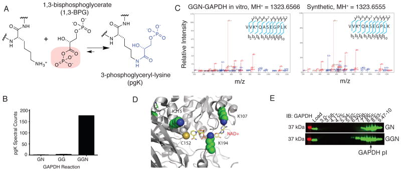
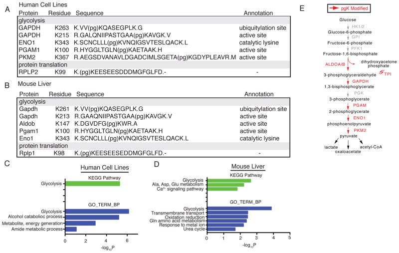
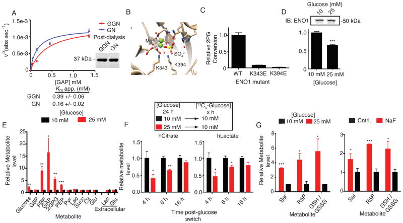
The posttranslational modification of proteins and their regulation by metabolites represent conserved mechanisms in biology. At the confluence of these two processes, we report that the primary glycolytic intermediate 1,3-bisphosphoglycerate (1,3-BPG) reacts with select lysine residues in proteins to form 3-phosphoglyceryl-lysine (pgK). This reaction, which does not require enzyme catalysis, but rather exploits the electrophilicity of 1,3-BPG, was found by proteomic profiling to be enriched on diverse classes of proteins and prominently in or around the active sites of glycolytic enzymes. pgK modifications inhibit glycolytic enzymes and, in cells exposed to high glucose, accumulate on these enzymes to create a potential feedback mechanism that contributes to the buildup and redirection of glycolytic intermediates to alternate biosynthetic pathways.


Figures

Fig. 1
Fig. 1
1,3-BPG forms a stable, covalent modification on lysines of GAPDH in vitro. (A) 3-phosphoglyceryl-lysine (pgK) formed by reaction of a lysine ε-amine with the acylphosphate functionality in 1,3-BPG. (B) Spectral counts of pgK-modified tryptic peptides detected by LC-MS/MS analyses of GN, GG, and GGN GAPDH enzymatic reactions (average of two independent experiments). (C) MS/MS spectra of the doubly charged synthetic (right) and in vitro GGN-GAPDH-derived (left) tryptic peptide VV(pg)KQASEGPLK. Observed b-, y-, and relevant parent ions, as well as products of dehydration (°) or ammonia loss (*) are labeled. “*” within peptide sequences denotes the pgK-modified lysine. (D) The most frequently detected pgK-modification sites (K107, K194 and K215) surround the active site of GAPDH (PDB Accession 1ZNQ). (E) α-GAPDH western blot of GG- and GGN-GAPDH reactions after IEF analysis. Data are from a representative experiment of three independent experiments.
Fig. 2
Fig. 2
Functional distribution of pgK modification sites in human cells and mouse tissues. (A, B) Modification site, peptide sequence, and associated annotation for representative endogenous pgK-modified proteins from human cell lines (A) and mouse liver (B). “(pg)K” denotes the pgK-modified lysine. (C, D) Gene ontology biological process categories (GOTERM_BP) and KEGG pathways enriched among pgK-modified proteins in human cell lines (C) and mouse liver (D) by DAVID bioinformatic analysis. (E) Schematic of observed pgK-modified enzymes in glycolysis. Glycolytic enzymes containing at least one pgK-site are shown in red, others are shown in grey.
Fig. 3
Fig. 3
Dynamic coupling of pgK modification to glucose metabolism. (A) Intracellular glucose and bisphosphoglycerate (BPG, aggregate of both 1,3- and 2,3-isomers) levels from cells grown at indicated glucose concentrations for 24 hours. (B) α-pgK immunoblot (IB) and Coomassie-stained gel of proteomes from HEK293T cells grown at indicated glucose concentrations. (C–D) α-pgK IB of α-FLAG-enriched ENO1 (C) and GAPDH (D) expressed in HEK293T cells grown at indicated glucose concentrations. Shown below each blot is a graph of the average relative α-pgK band intensities (n = 4 per group). (E) Representative SILAC chromatograms, MS1 isotope envelopes, and area ratios for non-pgK (left) and pgK-modified (right) peptides from ENO1. Integration area is shown within green bars; asterisk (*) in the chromatogram signifies the triggered MS2 scan. (F) Average SILAC ratios for pgK343-containing and non-pgK ENO1 peptides from cells grown at indicated glucose concentrations. Horizontal line and whiskers represent the mean and 10–90% confidence interval, respectively. (G) IB of IEF-focused ENO1 from MCF7 cells treated grown at indicated glucose concentrations. Plot shows the IEF-focused ENO1 pI distributions quantified by densitometry. (H) Spectral count values for pgK-modified Eno peptides across nine mouse tissues (Table S3). Data represent mean ± s.e.m; statistical significance was determined by two-way t-tests with a Bonferroni correction for C & D and Welch’s correction for F: *, P < 0.05; **, P < 0.01; ***, P < 0.005.
Fig. 4
Fig. 4
pgK modification impairs glycolytic enzymes and correlates with altered glycolytic output in human cells. (A) Michaelis-Menten kinetic analysis comparing GAPDH from GGN (1,3-BPG-producing) versus GN (control) reactions. (B) Structure of ENO1 active site (PDB Accession: 3B97) showing residues important for catalysis, including pgK sites K343 and K394. (C) Relative activities of wild-type and mutant FLAG-tagged ENO1 expressed and affinity-isolated from HEK293T cells (Fig. S17A for expression data of ENO1 variants). (D) Relative activities of FLAG-isolated ENO1 expressed in HEK293T cells cultured in 10 versus 25 mM glucose for 24 hours. (E) Relative metabolite levels in HEK293T cells grown in 10 versus 25 mM glucose for 24 hours. (F) Relative heavy lactate and citrate levels in HEK293T cells pretreated with the indicated glucose concentrations for 24 hours and then grown in 10 mM heavy-glucose for the indicated length of time. (G) Relative metabolite measurements in cells grown in 10 versus 25 mM glucose (left) or treated with NaF (right). Data shown represent mean ± s.e.m. from triplicate experiments. Statistical significance was determined by two-way t-tests: *, P < 0.05; **, P < 0.01; ***, P < 0.005; n.s., not significant. G6P, glucose-6-phosphate; FBP, fructose-1,6-bisphosphate; DHAP, dihydroxyacetonephosphate; Pyr, pyruvate; Lac, lactate; Succ, succinate; Glu, glutamate; Ser, serine; R5P, ribulose-5-phosphate; GSH/GSSG, reduced/oxidized glutathione.

References

    1. Swint-Kruse L, Matthews KS. Allostery in the LacI/GalR family: variations on a theme. Current opinion in microbiology. 2009 Apr;12:129. - PMC - PubMed
    1. Sola-Penna M, Da Silva D, Coelho WS, Marinho-Carvalho MM, Zancan P. Regulation of mammalian muscle type 6-phosphofructo-1-kinase and its implication for the control of the metabolism. IUBMB life. 2010 Nov;62:791. - PubMed
    1. Walsh C. Posttranslational modification of proteins : expanding nature’s inventory. Roberts and Co. Publishers; Englewood, Colo: 2006. p. xxi.p. 490.
    1. Wang T, Kartika R, Spiegel DA. Exploring post-translational arginine modification using chemically synthesized methylglyoxal hydroimidazolones. Journal of the American Chemical Society. 2012 May 30;134:8958. - PMC - PubMed
    1. Codreanu SG, Zhang B, Sobecki SM, Billheimer DD, Liebler DC. Global analysis of protein damage by the lipid electrophile 4-hydroxy-2-nonenal. Molecular & cellular proteomics : MCP. 2009 Apr;8:670. - PMC - PubMed

Publication types

MeSH terms