. 2013 Sep 27;113(8):965-74.
doi: 10.1161/CIRCRESAHA.113.301934. Epub 2013 Aug 1.

Increased atherosclerotic lesion formation and vascular leukocyte accumulation in renal impairment are mediated by interleukin-17A

Affiliations

Increased atherosclerotic lesion formation and vascular leukocyte accumulation in renal impairment are mediated by interleukin-17A

Shuwang Ge et al. Circ Res. .

Abstract

Rationale: Atherosclerosis is a major cause of death in patients with chronic kidney disease. Chronic inflammation of the arterial wall including invasion, proliferation, and differentiation of leukocytes is important in atherosclerotic lesion development. How atherosclerotic inflammation is altered in renal impairment is incompletely understood.

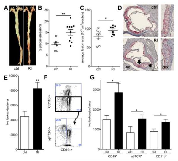
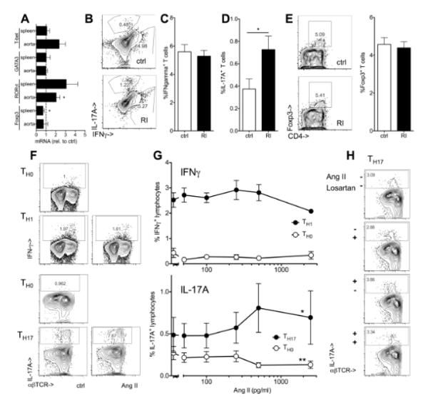
Objective: This study analyzed leukocytes of the atherosclerotic aorta in mice with impaired and normal renal function and studied a mechanism for the alteration in aortic myeloid leukocytes.

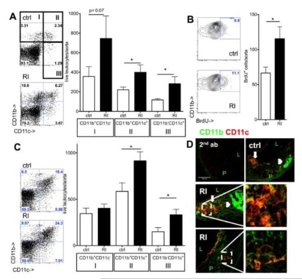
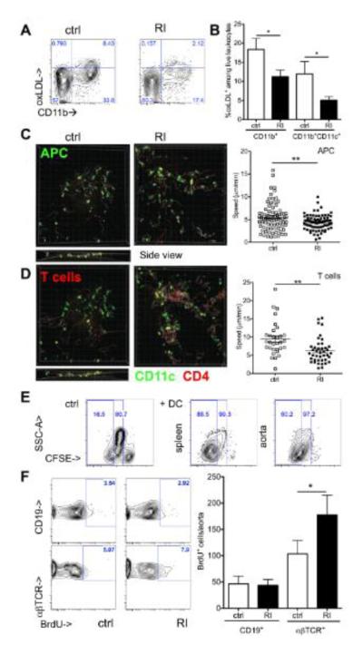
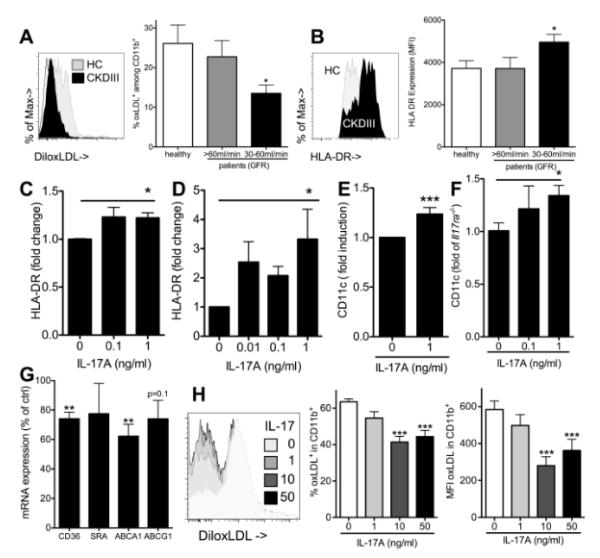
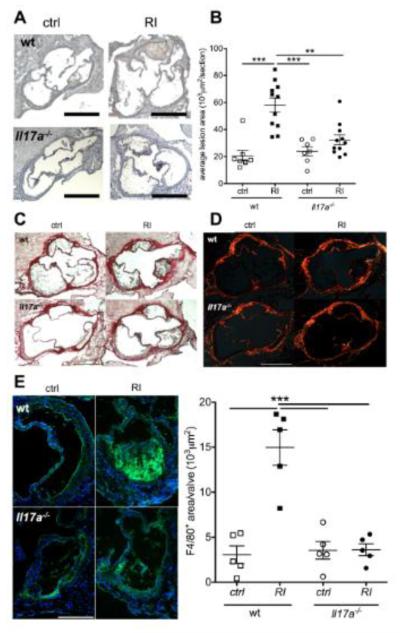
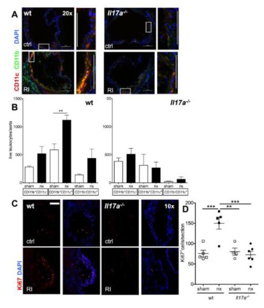
Methods and results: Unilateral nephrectomy significantly decreased glomerular filtration rate and increased atherosclerotic lesion size and aortic leukocyte numbers in 2 murine atherosclerosis models, apolipoprotein E (Apoe(-/-)) and low-density lipoprotein (LDL) receptor-deficient (LDLr(-/-)) mice. The number of aortic myeloid cells increased significantly. They took-up less oxidized LDL, whereas CD11c expression, interaction with T cells, and aortic T cell proliferation were significantly enhanced in renal impairment. In human peripheral blood mononuclear cell cultures, chronic kidney disease serum decreased lipid uptake and increased human leukocyte antigen II (HLA II) expression. Supplementation with interleukin-17A similarly increased HLA II and CD11c expression and impaired oxidized LDL uptake. Interleukin-17A expression was increased in atherosclerotic mice with renal impairment. Ablation of interleukin-17A in LDLr(-/-) mice by lethal irradiation and reconstitution with Il17a(-/-) bone marrow abolished the effect of renal impairment on aortic CD11b(+) myeloid cell accumulation, CD11c expression, and cell proliferation. Atherosclerotic lesion size was decreased to levels observed in normal kidney function.

Conclusions: Kidney function modifies arterial myeloid cell accumulation and phenotype in atherosclerosis. Our results suggest a central role for interleukin-17A in aggravation of vascular inflammation and atherosclerosis in renal impairment.

Keywords: atherosclerosis; interleukin-17; leukocytes; renal insufficiency; vascular inflammation.


Figures

Figure 1
Figure 1. Renal impairment increases atherosclerotic lesion size and leukocyte infiltration
(A,B) En face aortic atherosclerotic lesion size was assessed in Apoe−/− mice (12 weeks high fat diet) (A, ctrl: sham operated, RI, renal impairment) and quantified as percent of aortic surface area (B)(n=6-10, 3 independent experiments). (C,D) Aortic root lesion size in the 300 μm following the aortic valve (C, n=8, 4 independent experiments, D examples, 4x and 20x original magnification, arrows mark the same areas as in figure 2). (E-G) Aortic leukocyte numbers were analyzed by flow-cytometry (gated for live, CD45+ cells; E, n=9-11, 4 independent experiments). Staining for CD11b (myeloid cells), CD19 (B cells) and αβTCR (T cells) (example in F), was used for quantification of these subgroups (G, n=5-11 per group, 4 independent experiments).
Figure 2
Figure 2. Myeloid cell accumulation in the aorta during atherosclerosis development in renal impairment
(A) CD11b and CD11c expression on aortic leukocytes in Apoe−/− mice after three weeks high fat diet by flow cytometric analysis (ctrl=sham-operated, RI=renal impairment, examples and n=11-12, 4 independent experiments). (B) Proliferation of aortic myeloid cells by BrdU incorporation at the same timepoint (cell number/aorta, n=8-10 from 3 independent experiments). (C) Aortic flow cytometry after twelve weeks high fat diet showed increased CD11b+CD11c+ and CD11c+ cell numbers in renal impairment (n=9-11, 4 independent experiments). (D) Localization in established atherosclerotic lesions (12 weeks high fat diet) by confocal imaging revealed CD11c+ (red) cells in the neo-intima. Most of these cells were also CD11b+ (green) in both control and renal impairment (RI) Apoe−/− mice. CD11b+CD11c+ cells were present in foam cell (thick arrows; compare to lipid (figure 1D)) and highly cellular regions. CD11b was also found in a-cellular intimal areas (arrowheads compare to fig. 1D) (secondary antibodies only as negative control, P=plaque, L=lumen, 40x original magnification).
Figure 3
Figure 3. Altered lipid uptake and interaction with T cells of aortic myeloid leukocytes in renal impairment
(A,B) Uptake of DiI-oxLDL by aortic leukocytes was assessed 24h after injection into Apoe−/− mice with and without renal impairment (RI) by flow cytometry (A shown in all live leukocytes, B quantified as proportion of CD11b+ and CD11b+CD11c+ cells, n=4-7, 2 independent experiments). (C,D) Aortic antigen presentation was assessed by live cell 2-photon microscopy after co-incubation of explanted aortas from CD11cYFPApoe−/− mice (ctrl and RI) with SNARF labeled Apoe−/− CD4+ T cells (red). CD11c+ antigen presenting cell (APC, C) and T cell (D) velocities were assessed in control and RI aortas in three dimensions (z axis shown in ctrl, stills from suppl. movies 1-4, available online, average cell velocities given, 1 of 2 independent experiments, unpaired t-tests). (E) Proliferation of BALB/c lymphocytes in mixed lymphocyte reaction with splenic and aortic CD11b+CD11c+ cells from atherosclerotic Apoe−/− mice was assessed by CFSE dilution (1 of 2 independent experiments with similar results, 4-5 aorta donors per group). (F) Proliferation of Apoe−/− aortic lymphocytes after three weeks high fat diet (BrdU+ cells/aorta, n=8-10 from 3 independent experiments).
Figure 4
Figure 4. Renal impairment and IL-17A enhance antigen presenting cell marker expression and decrease oxLDL uptake in myeloid cell differentiation
(A) oxLDL uptake by human PBMC derived macrophages differentiated in the presence of serum from patients with stable chronic kidney disease (GFR above 60 ml/min=CKDI,II, n=5, GFR 30-60ml/min=CKDIII, n=11 and healthy controls (HC), n=9, 3 independent experiments, Bonferroni after One-way-ANOVA) (B) HLAII surface expression on human PBMC derived macrophages assessed by flow cytometry (CKDI,II, n=11, CKDIII, n=19, healthy controls, n=8, 6 independent experiments, Bonferroni after One-way-ANOVA). (C,D) HLAII expression on PBMC derived macrophages cultured with and without recombinant IL-17A (C,n=5, linear trend after One-way-ANOVA) and dendritic cells (D, promoted by culture with GM-CSF and IL-4, n=4, linear trend after One-way-ANOVA). (E,F) Murine macrophages were generated by culture of adherent bone marrow cells. The effect of recombinant mouse IL-17A on CD11c expression was investigated in both wt (E, 4 independent experiments) and Il17a−/− compared to Il17ra−/− mice (F, t-test after One-way-ANOVA, 3 independent experiments). (G) Il17a−/− macrophage mRNA expression was assessed on day three of culture in the presence of recombinant IL-17A (1ng/ml) and compared to control cells (t-tests, 3 independent experiments). (H) Wt macrophage oxLDL uptake on day 7 after culture with different concentrations of recombinant IL-17A (Dunnetts after One-way-ANOVA, n=12, 3 independent experiments).
Figure 5
Figure 5. Renal impairment and Angiotensin II enhance TH17 polarization
(A) mRNA expression of markers of T cell lineages (T-bet for the TH1, GATA-3 for TH2, RORγt for TH17 and Foxp3 for Treg) was assessed in spleens and aortas of Apoe−/− mice after 12 weeks high fat diet (renal impairment relative to controls, n=5, t-tests). (D-E) Interferon gamma (IFNγ)(B,C, n=6-8) and IL-17A producing (B,D, n=8-13) T cells and Foxp3+ regulatory T cells (E, n=4-5) in spleens from Apoe−/− mice with normal and impaired renal function (RI) (5h PMA/ionomycin, % of CD3+ cells, Il17a−/− spleens used to define IL-17A positivity (<0.1%)). (F,G) Mouse splenocytes were cultured under TH0, TH1 and TH17 polarizing conditions with and without exogenous Angiotensin II (AngII, 250 pg/ml unless indicated) and intracellular cytokine expression was assessed after re-stimulation as described in methods (F, typical examples and G, n=3 independent experiments, One-way ANOVA, *indicates significant slope). (H) TH17 polarization was carried out in the presence or absence of Angiotensin II and Losartan (1nM) or solvent control (one of 2 independent experiments).
Figure 6
Figure 6. IL-17A ablation abolishes atherosclerosis enhancement in renal impairment
Atherosclerosis development was studied in LDLr−/− mice reconstituted with either wt or Il17a−/− bone marrow and normal and impaired renal function. (A,B) Aortic root lesion size (A, examples, 5x orig. magn., bars=500μm, B means±SEM, n=7-11/group, 4 independent experiments, Bonferroni after One-way-ANOVA. (C,D) Picrosirius-red stain of collagen contents as translucent image (D) and with the use of polarized light (E, examples of at least 4 mice/group). (F) F4/80-macrophage staining (examples and statistical analysis of n=5 mice per group, F4/80 positive area/valvular lesion, Bonferroni after One-way-ANOVA).
Figure 7
Figure 7. IL-17A ablation abolishes enhanced aortic macrophage accumulation in renal impairment
The aortic leukocyte infiltrate was analyzed in LDLr−/− mice reconstituted with either wt or Il17a−/− bone marrow and normal and impaired renal function. (A) Immunofluorescence to assess CD11b (green) and CD11c (red) in the aortic root (typical examples of aortic valves, rectangles mark the cell rich intimal regions shown in zoom. 20x orig. magn., bars=100μm). (B) Number of total aortic CD11b+ and CD11c+ cells determined by flow cytometry (n=5-8, 4 independent experiments). (C,D) Aortic cell proliferation was assessed by Ki67 staining (E, examples of aortic valve area plaques, 10x orig. magn., bar=200μm, F: mean Ki67+ cell numbers/aortic section from n=4 sections/mouse, n=4-6 mice/group, One-way-ANOVA and Bonferroni post-hoc test).

Similar articles

Cited by

References

    1. Tonelli M, Wiebe N, Culleton B, House A, Rabbat C, Fok M, McAlister F, Garg AX. Chronic kidney disease and mortality risk: A systematic review. Journal of the American Society of Nephrology : JASN. 2006;17:2034–2047. - PubMed
    1. Astor BC, Hallan SI, Miller ER, 3rd, Yeung E, Coresh J. Glomerular filtration rate, albuminuria, and risk of cardiovascular and all-cause mortality in the us population. American journal of epidemiology. 2008;167:1226–1234. - PubMed
    1. Go AS, Chertow GM, Fan D, McCulloch CE, Hsu CY. Chronic kidney disease and the risks of death, cardiovascular events, and hospitalization. The New England journal of medicine. 2004;351:1296–1305. - PubMed
    1. Herzog CA, Asinger RW, Berger AK, Charytan DM, Diez J, Hart RG, Eckardt KU, Kasiske BL, McCullough PA, Passman RS, DeLoach SS, Pun PH, Ritz E. Cardiovascular disease in chronic kidney disease. A clinical update from kidney disease: Improving global outcomes (kdigo) Kidney Int. 2011;80:572–586. - PubMed
    1. Schiffrin EL, Lipman ML, Mann JF. Chronic kidney disease: Effects on the cardiovascular system. Circulation. 2007;116:85–97. - PubMed

Publication types

MeSH terms