Review
. 2014 Sep 5;9(9):1627-38.
doi: 10.2215/CJN.10391012. Epub 2013 Aug 1.

Proximal tubule function and response to acidosis

Affiliations
Review

Proximal tubule function and response to acidosis

Norman P Curthoys et al. Clin J Am Soc Nephrol. .

Abstract

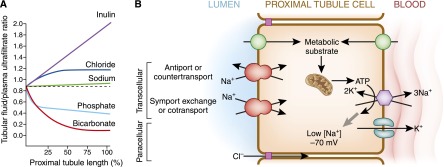
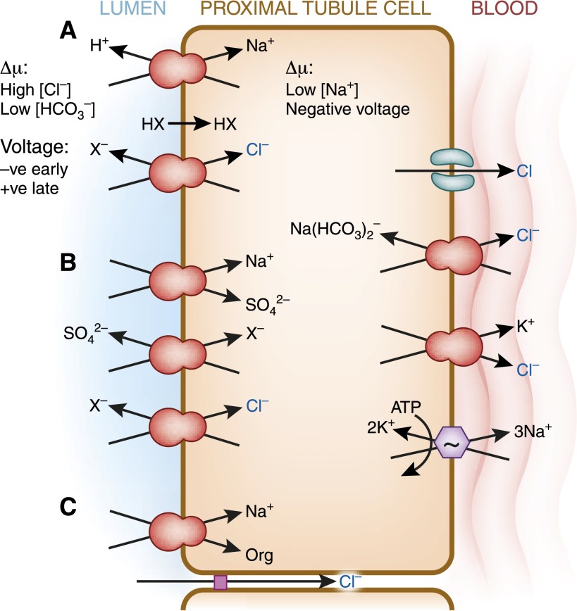
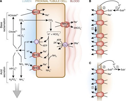
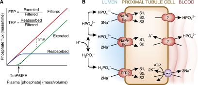
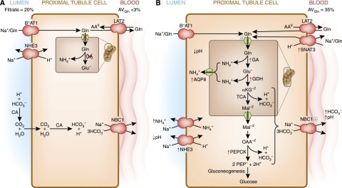
The human kidneys produce approximately 160-170 L of ultrafiltrate per day. The proximal tubule contributes to fluid, electrolyte, and nutrient homeostasis by reabsorbing approximately 60%-70% of the water and NaCl, a greater proportion of the NaHCO3, and nearly all of the nutrients in the ultrafiltrate. The proximal tubule is also the site of active solute secretion, hormone production, and many of the metabolic functions of the kidney. This review discusses the transport of NaCl, NaHCO3, glucose, amino acids, and two clinically important anions, citrate and phosphate. NaCl and the accompanying water are reabsorbed in an isotonic fashion. The energy that drives this process is generated largely by the basolateral Na(+)/K(+)-ATPase, which creates an inward negative membrane potential and Na(+)-gradient. Various Na(+)-dependent countertransporters and cotransporters use the energy of this gradient to promote the uptake of HCO3 (-) and various solutes, respectively. A Na(+)-dependent cotransporter mediates the movement of HCO3 (-) across the basolateral membrane, whereas various Na(+)-independent passive transporters accomplish the export of various other solutes. To illustrate its homeostatic feat, the proximal tubule alters its metabolism and transport properties in response to metabolic acidosis. The uptake and catabolism of glutamine and citrate are increased during acidosis, whereas the recovery of phosphate from the ultrafiltrate is decreased. The increased catabolism of glutamine results in increased ammoniagenesis and gluconeogenesis. Excretion of the resulting ammonium ions facilitates the excretion of acid, whereas the combined pathways accomplish the net production of HCO3 (-) ions that are added to the plasma to partially restore acid-base balance.


Figures

Figure 1.
Figure 1.
General considerations of proximal tubule transport. (A) Profile of the tubular fluid to plasma ultrafiltrate ratio (TF/PUF). Selected solutes are shown along the length of the proximal tubule. PUF is a surrogate for the proto-urine in Bowman’s space. Inulin represents a filtered molecule that is neither secreted nor reabsorbed and the rise in its TF/PUF solely reflects reabsorption of water, which concentrates luminal inulin. Sodium reabsorption is near isotonic with water, which results in a very small increase in TF/PUF by the end of the proximal tubule. HCO3 absorption in the early proximal convoluted tubule is particularly avid leading to rapid fall in TF/PUF. The fall in luminal [HCO3] is accompanied by a reciprocal rise in luminal [Cl] as reabsorption remains by-and-large isotonic. Inorganic phosphate (Pi) reabsorption is more avid in the earlier parts of the proximal tubule. (B) Generic scheme of the proximal tubule cell. The primary energy currency is organic metabolic substrates that enter the proximal tubule and are catabolized to produce ATP, which serves as the secondary energy currency. Some transporters are directly coupled to ATP hydrolysis (enthalpic transport), such as the H+-ATPase and Na+/K+-ATPase. The latter represents the main workhorse of the proximal tubule responsible for the majority of the cellular ATP consumption. The Na+/K+-ATPase converts the energy stored in ATP into low cellular [Na+] and high cellular [K+]. The presence of K+ conductance allows the [K+] gradient to increase the negative interior potential. The low cell [Na+] and negative voltage serve as the tertiary energy currencies that drive multiple secondary active apical transporters to achieve uphill movement of solutes coupled to downhill movement of Na+ (entropic transport). The transported solutes move in the same (symport or cotransport) or opposite (antiport, exchanger, or countertransport) direction as Na+. Movement of solute can also proceed via paracellular routes driven by electrochemical forces.
Figure 2.
Figure 2.
Proximal tubule NaCl reabsorption. Unlike the thick ascending limb or distal convoluted tubule, there are no dedicated “NaCl” transporters in the proximal tubule. The proximal tubule uses parallel arrays of symporters and antiporters to affect NaCl entry. (A) Parallel Na+/H+ exchange (NHE3) and Cl/base exchange (CFEX, SLC26A6) with recycling of the conjugate acid HX resulting in net NaCl entry. (B) A triple coupling mechanism where Na-sulfate cotransport (NaS-1) runs in parallel with two anion exchangers to achieve NaCl cotransport. (C) Na+-coupled organic solute transport with concurrent paracellular Cl transport driven by the lumen-to-interstitial space downhill chemical gradient of [Cl]. Basolateral Na+ exit is mediated by the Na+/K+-ATPase but some Na+ also exit with HCO3 (see Figure 3). Cl exit is less well defined utilizing a variety of transporters and possibly a Cl channel.
Figure 3.
Figure 3.
Proximal tubule HCO3 reabsorption and H+ secretion. Unlike NaCl reabsorption, luminal acidification is mediated by dedicated acid-base transporters. (A) Luminal H+ secretion is mediated by direct coupling to ATP hydrolysis via the H+-ATPase, but a higher proportion of luminal H+ secretion occurs by the Na+/H+ exchanger NHE3. In the neonate before maturational expression of NHE3, NHE8 is the more important NHE isoform. In addition to H+, NHE3 also directly transports NH4+ formed in the cell from NH3 and H+ into the lumen. The H+ secreted into the lumen has several fates. Reclamation of filtered base is shown on the top left. The titration of filtered HCO3 leads to formation of CO2 under the influence of carbonic anhydrase (CA). The CO2 enters the cell and is tantamount to HCO3 reabsorption. The titration of trivalent citrate to its bivalent form facilitates citrate reabsorption. Citrate is the most important and abundant organic urinary base equivalent. The bottom left depicts acid secretion. In addition to direct Na+/NH4+ exchange by NHE3, luminal H+ can combine with NH3 to form de novo NH4+ in the lumen. Titration of divalent to monovalent phosphate reduces phosphate absorption and allows the titrated phosphate to function as a H+ carrier in the urine. CO2 in the cell is hydrated to H2CO3, which dissociates to form a H+ and HCO3. The metabolism of citrate2−/3− also consumes H+ in the cell, a reaction equivalent to generating HCO3. The generated HCO3 exits the cell via Na+-coupled transport. There are three generic mechanisms by which changes in luminal and cell pH can alter apical transporters. (B and C) Stimulation is shown in B and inhibition is shown in C. (1) Luminal pH can alter the substrate concentration by titration and regulate transport kinetically. (2) Direct regulatory gating of the transporters by pH. (3) Changes in the number of transporters on the apical membrane by changes in trafficking, protein synthesis, and transcript levels. Sub, substrate.
Figure 4.
Figure 4.
Proximal tubule phosphate transport. (A) Concepts of renal inorganic phosphate (Pi) homeostasis by the proximal tubule. The flux of filtered and reabsorbed Pi is plotted against plasma phosphate concentration; the difference between the two yields the rate of excretion of Pi. There are a number of terms used to quantify the proximal tubule’s Pi reabsorption at the whole organism level. Fractional excretion of Pi (FEP) and tubular reabsorption of Pi (TRP) sum to unity (FEP=1−TRP). The maximal tubular reabsorptive capacity of Pi (TmP in units of mass/time) refers to the saturating transepithelial flux of Pi that the tubule can mount and is equal to the difference between filtered and absorbed phosphate when the filtered load is higher than TmP. The plasma concentration threshold at which Pi starts to appear in the urine is TmP/GFR (in units of mass/volume). (B) Cell model of proximal tubule Pi transport. Three apical transporters mediate Pi entry with different preferred valence of Pi, stoichiometry of Na+, electrogenicity, and pH gating. The affinities for Na+ are all approximately 30–50 mM but are much higher for phosphate (0.1, 0.07, and 0.025 mM for NaPi-Ila, NaPi-Ilc, and PiT-2, respectively). Distribution in the proximal tubule segments (S1, S2, S3). Basolateral Pi exit occurs via unknown mechanisms. Apical Na-coupled Pi transport is inhibited in acidosis by alteration in luminal substrate, directly gating of the transporter by pH, and decreased apical NaPi transporters as described in Figure 3B.
Figure 5.
Figure 5.
Renal proximal tubular catabolism of glutamine. (A) During normal acid-base balance, the glutamine filtered by the glomeruli is nearly quantitatively extracted from the lumen of the proximal convoluted tubule and largely returned to the blood. The transepithelial transport utilizes BoAT1, a Na+-dependent neutral amino acid cotransporter in the apical membrane, and LAT2, a neutral amino acid antiporter in the basolateral membrane. To accomplish this movement, either the mitochondrial glutamine transporter or the mitochondrial glutaminase (GA) must be inhibited (red X). The apical Na+/H+ exchanger functions to slightly acidify the lumen to facilitate the recovery of HCO3 ions. (B) During chronic acidosis, approximately one third of the plasma glutamine is extracted and catabolized within the early portion of the proximal tubule. BoAT1 continues to mediate the extraction of glutamine from the lumen. Uptake of glutamine through the basolateral membrane occurs by reversal of the neutral amino acid exchanger, LAT2, and through increased expression of SNAT3. Increased renal catabolism of glutamine is facilitated by increased expression (red arrows) of the genes that encode glutaminase (GA), glutamate dehydrogenase (GDH), phosphoenolpyruvate carboxykinase (PEPCK), the mitochondrial aquaporin-8 (AQP8), the apical Na+/H+ exchanger (NHE3), and the basolateral glutamine transporter (SNAT3). In addition, the activities of the mitochondrial glutamine transporter and the basolateral Na+/3HCO3 are increased (+). Increased expression of NHE3 contributes to the transport of ammonium ions and the acidification of the luminal fluid. The combined increases in renal ammonium ion excretion and gluconeogenesis result in a net synthesis of HCO3 ions that are transported across the basolateral membrane by the Na+/3HCO3 cotransporter (NBC1). CA, carbonic anhydrase; αKG, α-ketoglutarate; Mal, malate; OAA, oxaloacetate; PEP, phosphoenolpyruvate.

References

    1. Jones G, Prosser DE, Kaufmann M: 25-Hydroxyvitamin D-24-hydroxylase (CYP24A1): Its important role in the degradation of vitamin D. Arch Biochem Biophys 523: 9–18, 2012 - PubMed
    1. Meyer C, Dostou JM, Gerich JE: Role of the human kidney in glucose counterregulation. Diabetes 48: 943–948, 1999 - PubMed
    1. De Vito E, Cabrera RR, Fasciolo JC: Renin production and release by rat kidney slices. Am J Physiol 219: 1042–1045, 1970 - PubMed
    1. Koury ST, Bondurant MC, Koury MJ: Localization of erythropoietin synthesizing cells in murine kidneys by in situ hybridization. Blood 71: 524–527, 1988 - PubMed
    1. Moe OW, Ujiie K, Star RA, Miller RT, Widell J, Alpern RJ, Henrich WL: Renin expression in renal proximal tubule. J Clin Invest 91: 774–779, 1993 - PMC - PubMed

Publication types

MeSH terms