. 2013 Dec 1;208(11):1803-12.
doi: 10.1093/infdis/jit364. Epub 2013 Aug 1.

Suppression of PTRF alleviates the polymicrobial sepsis induced by cecal ligation and puncture in mice

Affiliations

Suppression of PTRF alleviates the polymicrobial sepsis induced by cecal ligation and puncture in mice

Yijie Zheng et al. J Infect Dis. .

Abstract

Background: Sepsis and sepsis-associated organ failure are devastating conditions. Understanding the detailed cellular/molecular mechanisms involved in sepsis should lead to the identification of novel therapeutic targets.

Methods: Cecal ligation and puncture (CLP) was used as a polymicrobial sepsis model in vivo to determine mortality and end-organ damage. Macrophages were adopted as the cellular model in vitro for mechanistic studies.

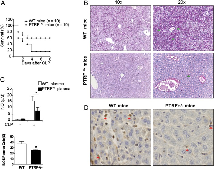
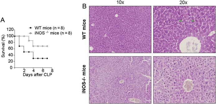
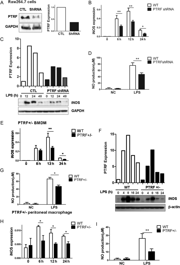
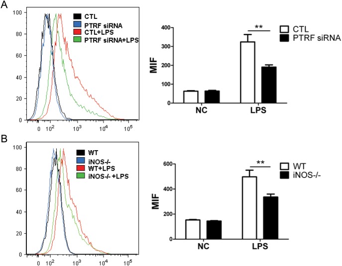
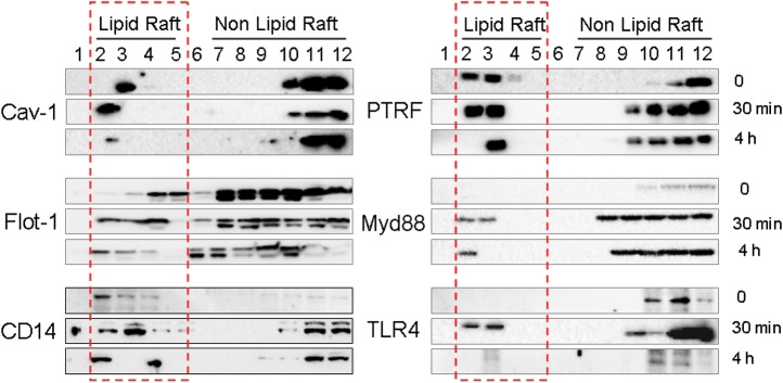
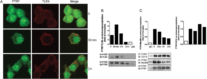
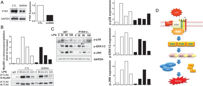
Results: PTRF+/- mice survived longer and suffered less organ damage after CLP. Reductions in nitric oxide (NO) and iNOS biosynthesis were observed in plasma, macrophages, and vital organs in the PTRF+/- mice. Using an acute sepsis model after CLP, we found that iNOS-/- mice had a comparable level of survival as the PTRF+/- mice. Similarly, polymerase I transcript release factor (PTRF) deficiency resulted in decreased iNOS and NO/ROS production in macrophages in vitro. Mechanistically, lipopolysaccharide (LPS) enhanced the co-localization and interaction between PTRF and TLR4 in lipid rafts. Deletion of PTRF blocked formation of the TLR4/Myd88 complex after LPS. Consistent with this, lack of PTRF impaired the TLR4 signaling, as shown by the decreased p-JNK, p-ERK, and p-p38, which are upstream factors involved in iNOS transcription.

Conclusion: PTRF is a crucial regulator of TLR4 signaling in the development of sepsis.

Keywords: CLP; PTRF; ROS; TLR4; macrophage; nitric oxide; sepsis.


Figures

Figure 1.
Figure 1.
Deletion of PTRF decreased iNOS expression in vivo, reduced mortality and organ damage in CLP-induced sepsis. A, PTRF+/− mice (open circles; n = 10) and WT mice (closed circles; n = 10) were subjected to CLP and monitored for survival. BE, Tissues and plasma were collected 24 hours after CLP. B, PAS staining was performed on liver tissue to evaluate acute hepatocyte injury. Light arrow: positive for PAS staining (C) NO level in plasma was measured using Griess reagent. D, Paraffin-embedded sections of liver tissue were obtained from WT mice (left panel) and PTRF+/− mice (right panel) after CLP. Liver tissue was stained for iNOS expression (dark arrow) using polyclonal anti-iNOS. E, iNOS positive cells were quantified in the liver sections obtained from WT mice or PTRF+/− mice. All figures above represented 3 independent experiments with similar results. *P < .05. Abbreviations: CLP, cecal ligation and puncture; PTRF, polymerase I transcript release factor; WT, wild type.
Figure 2.
Figure 2.
Deletion of iNOS reduced mortality and organ damage in CLP-induced sepsis. A, iNOS−/− mice (open circles; n = 8) and WT mice (closed circles; n = 8) were subjected to CLP and monitored for survival. B, Liver tissue was collected from WT and iNOS−/− mice 24 hours after CLP, and PAS stain was obtained. Arrow: positive for PAS staining. All figures represented 3 independent experiments with similar results. Abbreviations: CLP, cecal ligation and puncture; WT, wild type.
Figure 3.
Figure 3.
Deletion of PTRF inhibited LPS-induced iNOS expression and NO synthesis in macrophages. A, Raw264.7 cells were transfected with PTRF shRNA plasmid (ShRNA) and negative control plasmid (CTL). After 48 hours, the efficiency of deleting PTRF was assessed by western blot analysis. Densitometry was used to quantify the density of bands. BD, The transfected Raw264.7 cells were stimulated with LPS (200 ng/mL) for indicated time. B, iNOS mRNA was determined by real-time PCR and standardized to the endogenous control GAPDH. C, iNOS protein expression was determined using western blot analysis and standardized to the endogenous control GAPDH. D, LPS induced NO production was measured using the Greiss reagent in Raw 264.7 cells transfected with shRNA or control plasmid. EH, Bone marrow derived macrophages (BMDM) were obtained from PTRF+/− mice. Cells were then stimulated with LPS (200 ng/mL) for indicated time. E, iNOS mRNA transcription was measured by real-time PCR and standardized to the endogenous control GAPDH. F, iNOS protein expression was determined using western blot analysis and standardized to the endogenous control GAPDH. G, NO production was measured using the Griess reagent. H and I, PTRF+/− peritoneal macrophages were derived from PTRF+/− mice. Cells were then stimulated with LPS (200 ng/mL) for indicated time. H, iNOS mRNA was measured by real-time PCR and standardized to the endogenous control GAPDH. I, NO production was detected using the Griess reagent. All figures above represented three independent experiments with similar results. *P < .05; **P < .01. Abbreviations: LPS, lipopolysaccharide; mRNA, messenger RNA; NO, nitric oxide; PCR, polymerase chain reaction; PTRF, polymerase I transcript release factor.
Figure 4.
Figure 4.
Deletion of PTRF or iNOS reduced the LPS-induced ROS generation. A, Raw264.7 cells were transfected with PTRF siRNA or control siRNA (CTL). After 36 hours, cells were stimulated with LPS (200 ng/mL). After additional 6 hours, the production of total ROS was determined using flow cytometry. Mean fluorescence intensity (MFI) was analyzed by 3 independent experiments. B, BMDM was obtained from iNOS−/− mice. Cells were then stimulated with LPS (200 ng/mL). After 6 hours, the total ROS was measured as above. MFI was analyzed by three independent experiments. **P < .01. Abbreviations: BMDM, bone-marrow derived macrophages; LPS, lipopolysaccharide; PTRF, polymerase I transcript release factor; ROS, reactive oxygen species.
Figure 5.
Figure 5.
TLR4 signaling components trafficked into lipid rafts after LPS stimulation. Raw264.7 cells were treated with LPS (200 ng/mL). After 30 minutes and 4 hours respectively, lipid raft and non raft fractions were isolated as described in Material and Method. Fractions were then analyzed for cav-1, PTRF, Flot-1, Myd88, CD14, and TLR4 using Western blot analysis. Red frame: lipid raft portion. All figures represented 3 independent experiments with similar results. Abbreviations: LPS, lipopolysaccharide; PTRF, polymerase I transcript release factor.
Figure 6.
Figure 6.
PTRF co-localized and interacted with TLR4 complex. A, Raw264.7 cells were treated with LPS (200 ng/mL). After 30 minutes or 4 hours, cells were stained with anti-PTRF, anti-TLR4, and DAPI (not shown). Co-localization of PTRF and TLR4 was observed using confocal microscopy. Light cells: PTRF; Dark cells: TLR4; Areas indicated by arrows: merge, co-localization of PTRF and TLR4; (B) Raw264.7 cells were treated with LPS (200 ng/mL). After 30 minutes, 4 hours, and 24 hours, respectively, cell lysate was collected for co-IP assays. PTRF was precipitated with rabbit polyclonal anti-PTRF antibody. TLR4 and PTRF (input) levels were determined using western blot analysis. Density of the bands was quantified by densitometry. All figures above represented at least 3 independent experiments with similar results. C, PTRF interacted with Myd88 and CD14. Raw264.7 cells were treated with LPS (200 ng/mL). After 30 minutes, 4 hours, and 24 hours, respectively, cell lysate was collected for co-IP assays. Myd88, CD14, and PTRF (input) levels were determined using Western blot analysis. Density of the bands was quantified by densitometry. Abbreviations: co-IP, co-immunoprecipitation assay; LPS, lipopolysaccharide; PTRF, polymerase I transcript release factor.
Figure 7.
Figure 7.
Deletion of PTRF inhibited the activation of TLR4 signaling. A, Raw264.7 cells were transfected with PTRF ShRNA plasmid (ShRNA) and control plasmid (CTL). The transfection efficiency was assessed by Western blot analysis. B, Deletion of PTRF affected TLR4/Myd88 complex formation. Transfected cells were treated with LPS (200 ng/mL) for indicated time. Interactions between TLR4 and Myd88 in control cells and PTRF shRNA transfected cells were determined using co-IP assays. Cell lysates were subjected to immunoprecipitation (IP) with anti-TLR4 and blotted with anti-Myd88 or anti-TLR4 (input). C, Deficiency of PTRF affected TLR4 down-stream signaling. BMDMs were obtained from WT or PTRF+/− mice. Cells were then stimulated with LPS for indicated time. Cell lysates were blotted with anti-phospho-p38 (p-p38), anti-phospho-ERK (p-ERK), anti-phospho-JNK (p-JNK). The relative expressions were standardized to the endogenous control GAPDH. Density of bands was quantified using densitometry. All figures above represented at least three independent experiments with similar results. D, Schemata of proposed mechanisms. PTRF interacts with TLR4 and assists the TLR4/Myd88 complex formation in the presence of LPS stimulation thus, promotes LPS induced TLR4/Myd88 signaling. Subsequently, this complex activates the down-stream MARK pathways and MARK pathway associated iNOS transcription. These augmented iNOS expression and NO production, along with fluxed ROS release lead to cellular injury and tissue damage in CLP induced sepsis. When PTRF is deleted, the LPS induced TLR4/Myd88 interaction decreases. The activation of MARK pathway and its down-stream iNOS/NO production are impaired. Thus, deletion of PTRF confers a protective role in CLP/LPS induced cellular injury and tissue damage. Abbreviations: co-IP, co-immunoprecipitation assay; LPS, lipopolysaccharide; PTRF, polymerase I transcript release factor; ROS, reactive oxygen species.

Similar articles

Cited by

References

    1. Levy MM, Fink MP, Marshall JC, et al. 2001 Sccm/Esicm/Accp/Ats/Sis international sepsis definitions conference. Crit Care Med. 2003;31:1250–6. - PubMed
    1. Munford RS. Severe sepsis and septic shock: the role of gram-negative bacteremia. Annu Rev Pathol. 2006;1:467–96. - PubMed
    1. Annane D, Bellissant E, Cavaillon JM. Septic shock. Lancet. 2005;365:63–78. - PubMed
    1. Biswas SK, Chittezhath M, Shalova IN, Lim JY. Macrophage polarization and plasticity in health and disease. Immunol Res. 2012;53:11–24. - PubMed
    1. Murray PJ, Wynn TA. Protective and pathogenic functions of macrophage subsets. Nat Rev Immunol. 2011;11:723–37. - PMC - PubMed

Publication types

MeSH terms