. 2013 Aug;15(8):875-87.
doi: 10.1593/neo.13786.

Host matrix modulation by tumor exosomes promotes motility and invasiveness

Affiliations

Host matrix modulation by tumor exosomes promotes motility and invasiveness

Wei Mu et al. Neoplasia. 2013 Aug.

Abstract

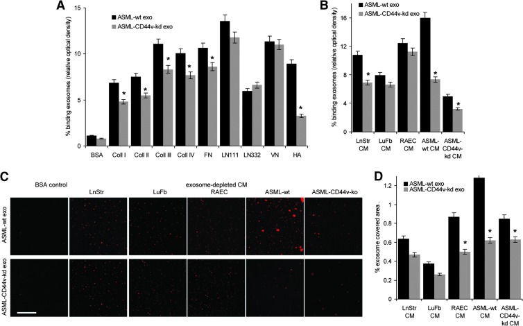
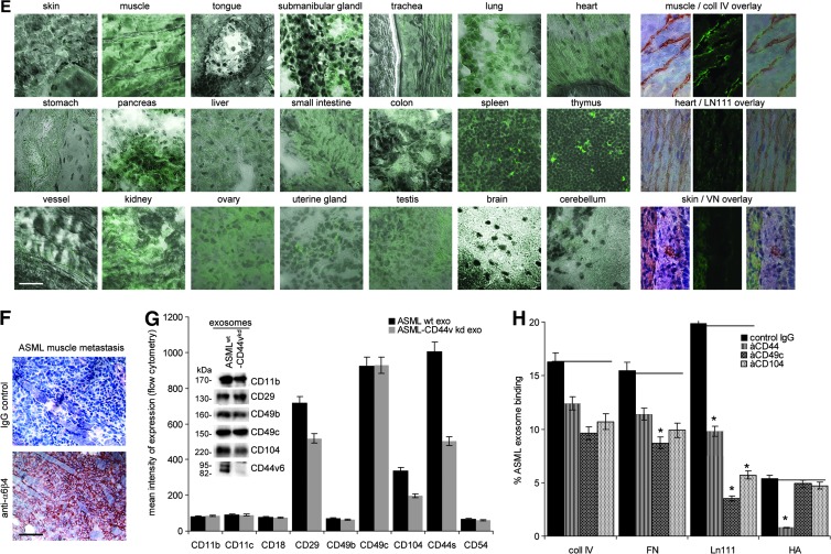
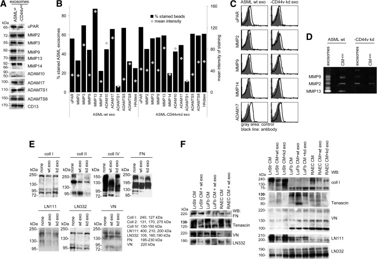
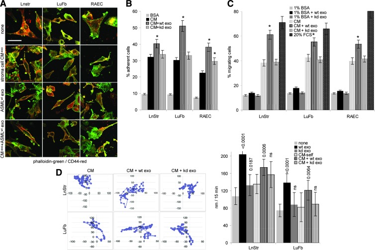
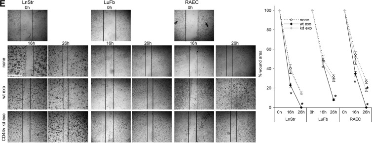
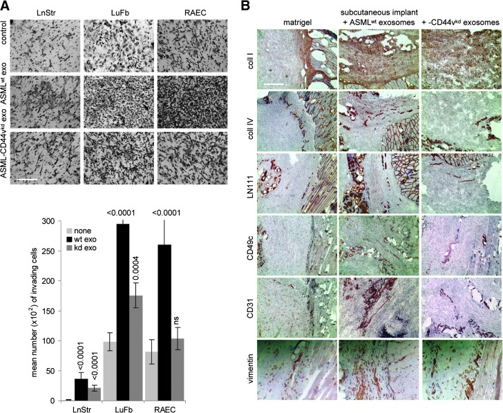
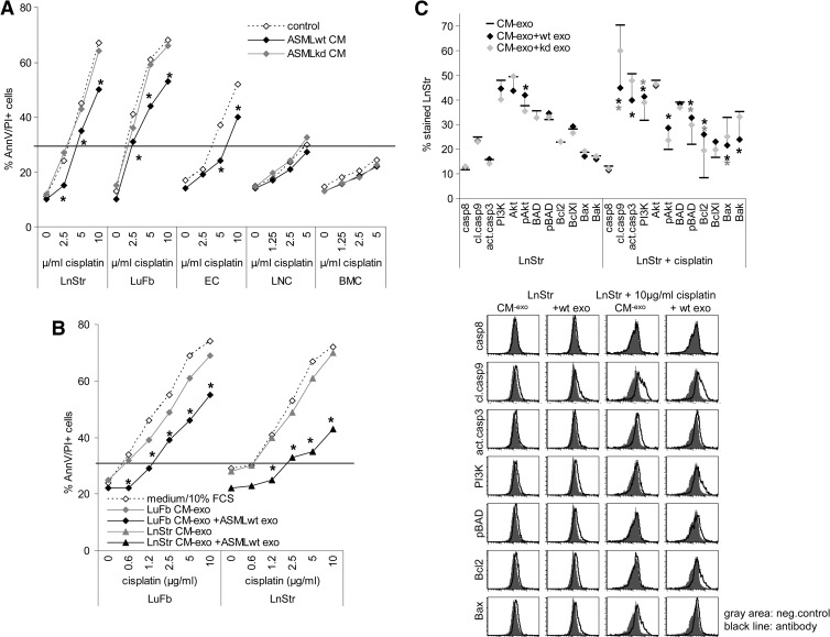
Exosomes are important intercellular communicators, where tumor exosomes (TEX) severely influence hematopoiesis and premetastatic organ cells. With the extracellular matrix (ECM) being an essential constituent of non-transformed tissues and tumors, we asked whether exosomes from a metastatic rat tumor also affect the organization of the ECM and whether this has consequences on host and tumor cell motility. TEX bind to individual components of the ECM, the preferential partner depending on the exosomes' adhesion molecule profile such that high CD44 expression is accompanied by hyaluronic acid binding and high α6β4 expression by laminin (LN) 332 binding, which findings were confirmed by antibody blocking. TEX can bind to the tumor matrix already during exosome delivery but also come in contact with distinct organ matrices. Being rich in proteases, TEX modulate the ECM as demonstrated for degradation of collagens, LNs, and fibronectin. Matrix degradation by TEX has severe consequences on tumor and host cell adhesion, motility, and invasiveness. By ECM degradation, TEX also promote host cell proliferation and apoptosis resistance. Taken together, the host tissue ECM modulation by TEX is an important factor in the cross talk between a tumor and the host including premetastatic niche preparation and the recruitment of hematopoietic cells. Reorganization of the ECM by exosomes likely also contributes to organogenesis, physiological and pathologic angiogenesis, wound healing, and clotting after vessel disruption.


Figures

Figure 1
Figure 1
Exosome binding to matrix proteins. (A–D) Dye-labeled ASMLwt and ASML-CD44vkd exosomes were incubated with (A) matrix protein-coated latex beads or (B) matrix protein- or (C and D) CM-exo-coated ELISA plates or glass slides. Exosome binding was evaluated by (A) flow cytometry, (B) OD, and (C and D) confocal microscopy. (A, B, and D) Mean values ± SD of triplicates and (C) representative examples are shown (scale bar, 10 µm). (E) Dye-labeled ASMLwt and ASML-CD44vkd exosomes (200 µg) were i.v. injected. Rats were sacrificed after 48 hours, and organs were excised and shock frozen. Tissue sections were counterstained with hematoxylin and eosin (H&E), evaluating recovery of exosomes by confocal microscopy. For selected samples, overlays with immunohistochemistry staining for matrix proteins are shown (scale bar, 10 µm). (F) BDX rats received ASML cells intrafootpad. Abdominal wall muscle was excised at autopsy and shock frozen. Tissue sections were stained with control IgG or B5.5 (anti-α6β4; scale bar, 20 µm). (G) Flow cytometry and WB analysis of adhesion molecule expression on ASMLwt and ASML-CD44vkd exosomes. (H) Dye-labeled exosomes were incubated with the indicated antibodies. Non-bound antibodies were removed by centrifugation (90 minutes, 100,000g). Exosomes (pellet) were collected and incubated with the indicated matrix proteins coated on ELISA plates. After 2 hours at 4°C, non-bound exosomes were removed by washing and fluorescence intensity (% of total exosomes) was evaluated. Means ± SD of triplicates are shown. Significant inhibition of exosome binding is indicated by an asterisk. Exosomes selectively bind matrix proteins and tissue matrices through adhesion receptors in vitro and in vivo, where TEX-decorated matrices appear to attract tumor cells.
Figure 1
Figure 1
Exosome binding to matrix proteins. (A–D) Dye-labeled ASMLwt and ASML-CD44vkd exosomes were incubated with (A) matrix protein-coated latex beads or (B) matrix protein- or (C and D) CM-exo-coated ELISA plates or glass slides. Exosome binding was evaluated by (A) flow cytometry, (B) OD, and (C and D) confocal microscopy. (A, B, and D) Mean values ± SD of triplicates and (C) representative examples are shown (scale bar, 10 µm). (E) Dye-labeled ASMLwt and ASML-CD44vkd exosomes (200 µg) were i.v. injected. Rats were sacrificed after 48 hours, and organs were excised and shock frozen. Tissue sections were counterstained with hematoxylin and eosin (H&E), evaluating recovery of exosomes by confocal microscopy. For selected samples, overlays with immunohistochemistry staining for matrix proteins are shown (scale bar, 10 µm). (F) BDX rats received ASML cells intrafootpad. Abdominal wall muscle was excised at autopsy and shock frozen. Tissue sections were stained with control IgG or B5.5 (anti-α6β4; scale bar, 20 µm). (G) Flow cytometry and WB analysis of adhesion molecule expression on ASMLwt and ASML-CD44vkd exosomes. (H) Dye-labeled exosomes were incubated with the indicated antibodies. Non-bound antibodies were removed by centrifugation (90 minutes, 100,000g). Exosomes (pellet) were collected and incubated with the indicated matrix proteins coated on ELISA plates. After 2 hours at 4°C, non-bound exosomes were removed by washing and fluorescence intensity (% of total exosomes) was evaluated. Means ± SD of triplicates are shown. Significant inhibition of exosome binding is indicated by an asterisk. Exosomes selectively bind matrix proteins and tissue matrices through adhesion receptors in vitro and in vivo, where TEX-decorated matrices appear to attract tumor cells.
Figure 2
Figure 2
Exosomes and matrix degradation. (A) Matrix-degrading enzymes in ASMLwt and ASML-CD44vkd exosomes were evaluated by WB and flow cytometry, (B) mean values of triplicates (percentage of stained beads and mean intensity of staining), and (C) representative examples. (D) Zymography of ASMLwt and ASML-CD44vkd CM-exo and exosomes. (E and F) WB of matrix proteins, LnStr-CM-exo, LuFb-CM-exo, and RAEC CM-exo after co-culture with ASMLwt and ASML-CD44vkd exosomes. Blots were incubated with the indicated antibodies. The expected size of matrix proteins and breakdown products is indicated. Coll II is degraded by ASMLwt and ASML-CD44vkd exosomes. Coll I, coll IV, FN, LN111, and LN332 are more efficiently degraded by ASMLwt than ASML-CD44vkd exosomes. This accounts for purified matrix proteins and the stroma cell matrix.
Figure 3
Figure 3
TEX-modulated CM and cell adhesion/motility. (A) Cells were seeded on cover slides coated with BSA, CM-exo, or ASMLwt exosome-treated CM-exo and were stained with phalloidin-fluorescein isothiocyanate (FITC) and anti-CD44-Cy3, where indicated cultures contained ASMLwt exosomes. Representative examples (confocal microscopy; scale bar, 100 µm) are shown. (B) LnStr, LuFb, and RAEC were seeded on BSA, CM-exo, or CM-exo plus ASMLwt or ASML-CD44vkd exosome-coated 96-well plates. Adhesion was evaluated after 2 hours (crystal violet staining of adherent cells). The mean ± SD of the percent adherent cells is shown. (C) Cells were seeded in the upper part of a Boyden chamber, and the lower part contained BSA, 20% FCS, or CM-exo pretreated with ASMLwt or ASML-CD44vkd exosomes as indicated. Migration was evaluated after 16 hours by crystal violet staining of the lower membrane site. Mean values (triplicates) ± SD of the percentage of migrating cells are shown. (D) Cells were seeded on CM-exo or TEX-pretreated CM-exo. Cell migration was observed for 24 hours. Representative examples and the mean ± SD track of 10 cells per 15 minutes are shown. (E) Cells were seeded on plates coated with CM-exo or TEX-pretreated CM-exo. Subconfluent monolayers were scratched with a pipette tip and wound closure was followed for 26 hours. Representative examples (scale bar, 250 µm) and mean ± SD (three wells) of wound closure are shown. (B-E) Significant differences between CM-exo and TEX-pretreated CM-exo are shown or indicated by asterisk. The TEX-modulated stroma matrix promotes stroma cell motility.
Figure 3
Figure 3
TEX-modulated CM and cell adhesion/motility. (A) Cells were seeded on cover slides coated with BSA, CM-exo, or ASMLwt exosome-treated CM-exo and were stained with phalloidin-fluorescein isothiocyanate (FITC) and anti-CD44-Cy3, where indicated cultures contained ASMLwt exosomes. Representative examples (confocal microscopy; scale bar, 100 µm) are shown. (B) LnStr, LuFb, and RAEC were seeded on BSA, CM-exo, or CM-exo plus ASMLwt or ASML-CD44vkd exosome-coated 96-well plates. Adhesion was evaluated after 2 hours (crystal violet staining of adherent cells). The mean ± SD of the percent adherent cells is shown. (C) Cells were seeded in the upper part of a Boyden chamber, and the lower part contained BSA, 20% FCS, or CM-exo pretreated with ASMLwt or ASML-CD44vkd exosomes as indicated. Migration was evaluated after 16 hours by crystal violet staining of the lower membrane site. Mean values (triplicates) ± SD of the percentage of migrating cells are shown. (D) Cells were seeded on CM-exo or TEX-pretreated CM-exo. Cell migration was observed for 24 hours. Representative examples and the mean ± SD track of 10 cells per 15 minutes are shown. (E) Cells were seeded on plates coated with CM-exo or TEX-pretreated CM-exo. Subconfluent monolayers were scratched with a pipette tip and wound closure was followed for 26 hours. Representative examples (scale bar, 250 µm) and mean ± SD (three wells) of wound closure are shown. (B-E) Significant differences between CM-exo and TEX-pretreated CM-exo are shown or indicated by asterisk. The TEX-modulated stroma matrix promotes stroma cell motility.
Figure 4
Figure 4
TEX-modulated CM promotes invasiveness. (A) Matrigel was mixed (1:1) with RPMI 1640 or TEX and was seeded on the lower membrane site of a transwell insert. LuFb, LnStr, and RAEC (5 x 10⁴) were layered on the upper site of the insert. Matrigel immigration was evaluated after 24 hours. Representative examples (scale bar, 200 µm) and mean numbers (triplicates) ± SD of matrigel invading cells are shown. Significant differences between matrigel and TEX-pretreated matrigel are indicated by asterisk. (B) Matrigel was mixed (1:1) with PBS, which contained ASMLwt or ASML-CD44vkd exosomes, as indicated. Matrigel was incubated for 12 hours at 37°C and was thereafter s.c. injected. The plug was removed after 5 days and was shock frozen. Plug sections were stained with anti-coll I, anti-coll IV, anti-LNγ1, anti-CD49c, anti-CD31, or anti-vimentin and were counterstained with H&E. Representative examples of the matrigel plug adjacent to host tissue are shown (scale bar, 200 µm). The exosome-modulated stroma matrix facilitates invasiveness.
Figure 5
Figure 5
TEX-modulated CM promotes hematopoietic and stroma cell proliferation. (A and B) Cells were incubated with CM, CM-exo, or TEX-pretreated CM-exo, where TEX were removed by centrifugation. Proliferative activity was evaluated by 3H-thymidine incorporation after 3 days of culture. (C) Flow cytometry analysis of LnStr and LuFb that were treated o/n with CM-exo with or without ASMLwt or ASML-CD44vkd exosomes. Mean values (three assays) of stained cells are shown. (D) LnStr and LuFb were incubated o/n with CM-exo or ASMLwt exosome- pretreated CM-exo, where TEX were removed by centrifugation. LnStr and LuFb were stained with the indicated antibodies, evaluating protein expression by flow cytometry. Representative examples and mean values (triplicates) are shown. (A-D) Significant differences between CM-exo and TEX-pretreated CM-exo are indicated by asterisk. (E) Evaluation of cytokines and chemokines in LnStr, LuFb, and RAEC CM-exo by WB. Growth promotion by TEX-treated LnStr and LuFb CM is accompanied by pronounced activation of the MAPK and JNK pathways and could be initiated by the liberation of growth factors from the CM by TEX, the LnStr, LuFb, and RAEC CM being rich in bFGF, HGF, SDF1, and TF.
Figure 6
Figure 6
TEX-modulated CM promotes stroma cell drug resistance. (A and B) Cells were incubated with ASML CM or TEX-pretreated CM-exo, removing TEX by centrifugation. Apoptosis resistance was evaluated by annexin V/propidium iodide (AnnV/PI) staining after 3 days of culture in the presence of titrated amounts of cisplatin. Mean values (triplicates) are shown. (C) Cells were incubated for 24 hours with CM-exo or TEX-pretreated CM-exo, where indicated cultures contained 10 µg/ml cisplatin. Cells were fixed and permeabilized, and expression of the indicated apoptosis/anti-apoptosis markers was evaluated by flow cytometry. Representative examples and mean values (three assays) are shown. (A–C) Significant differences between CM-exo and TEX-pretreated CM-exo are indicated by asterisk. Modulation of the stroma matrix by TEX promotes drug resistance of stroma cells, initiated by pronounced activation of the PI3K/Akt pathway.

References

    1. Hanahan D, Weinberg RA. Hallmarks of cancer: the next generation. Cell. 2011;144:646–674. - PubMed
    1. Hanahan D, Folkman J. Patterns and emerging mechanisms of the angiogenic switch during tumorigenesis. Cell. 1996;86:353–364. - PubMed
    1. Stewart TJ, Smyth MJ. Improving cancer immunotherapy by targeting tumor-induced immune suppression. Cancer Metastasis Rev. 2011;30:125–140. - PubMed
    1. Brabletz T. EMT and MET in metastasis: where are the cancer stem cells? Cancer Cell. 2012;22:699–701. - PubMed
    1. Kaplan RN, Rafii S, Lyden D. Preparing the “soil”: the premetastatic niche. Cancer Res. 2006;66:11089–11093. - PMC - PubMed

Publication types

MeSH terms