. 2013 Jul 30:7:25.
doi: 10.3389/fnana.2013.00025. eCollection 2013.

Noise-induced hearing loss is correlated with alterations in the expression of GABAB receptors and PKC gamma in the murine cochlear nucleus complex

Affiliations

Noise-induced hearing loss is correlated with alterations in the expression of GABAB receptors and PKC gamma in the murine cochlear nucleus complex

Zhen-Zhen Kou et al. Front Neuroanat. .

Abstract

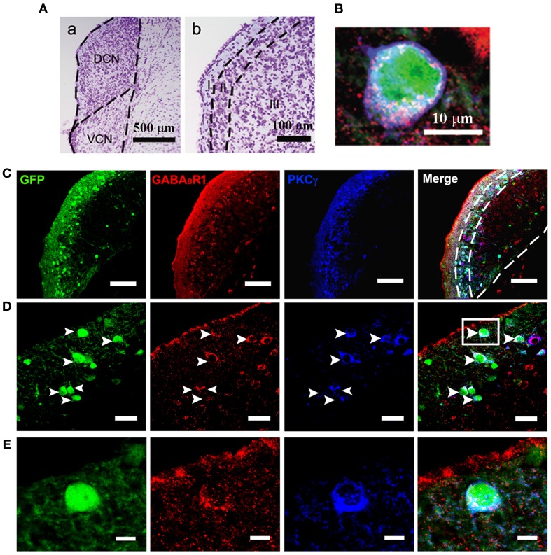
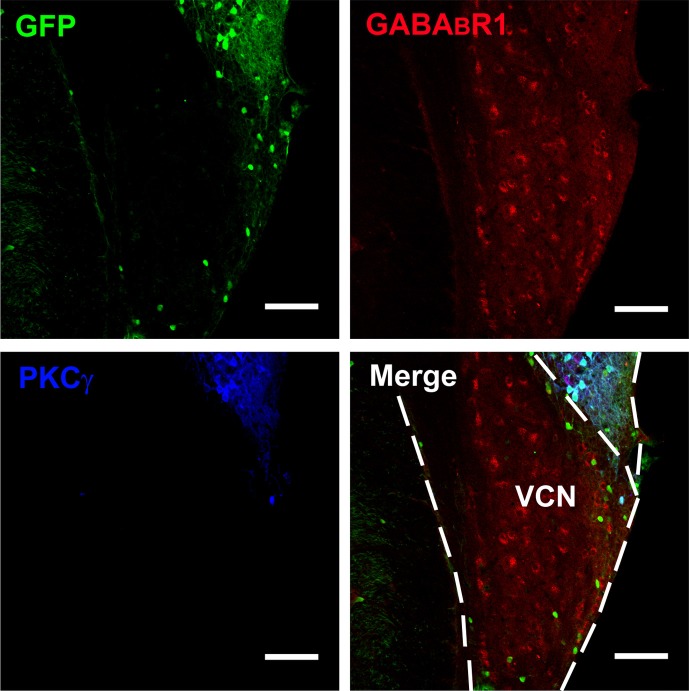
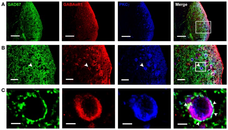
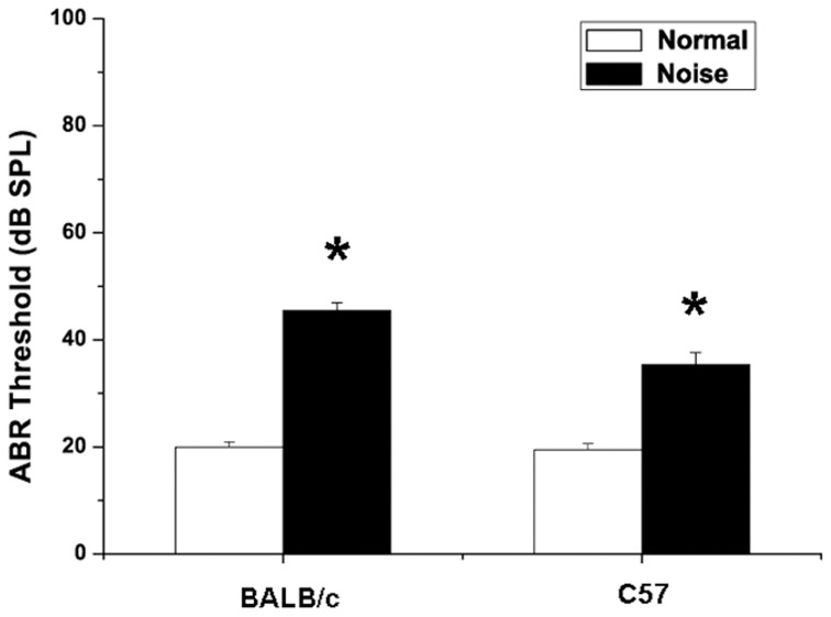
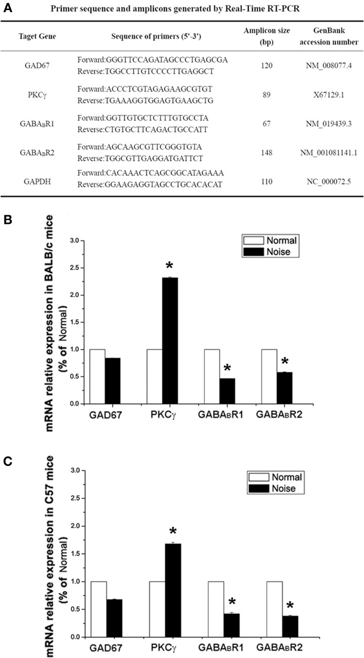
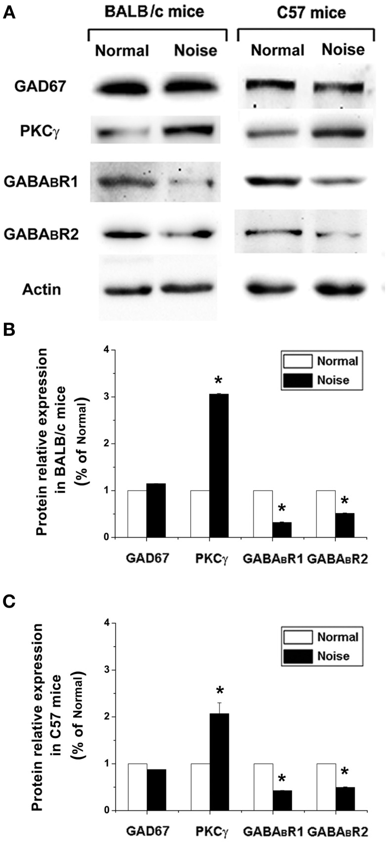
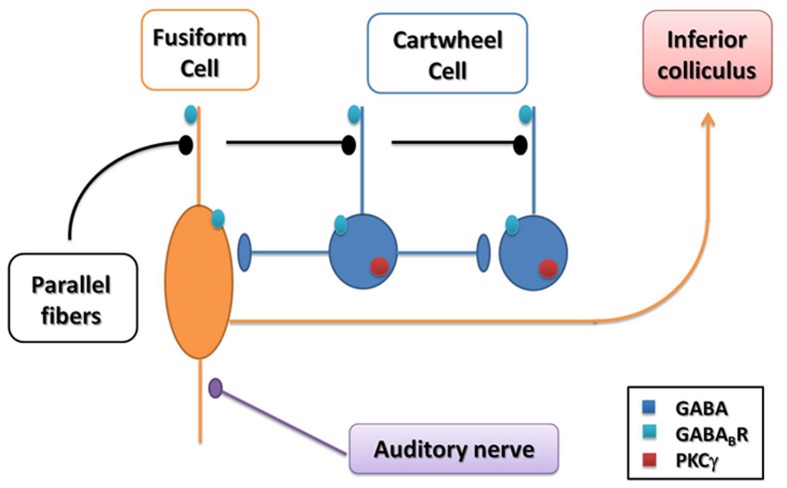
Noise overexposure may induce permanent noise-induced hearing loss (NIHL). The cochlear nucleus complex (CNC) is the entry point for sensory information in the central auditory system. Impairments in gamma-aminobutyric acid (GABA)-mediated synaptic transmission in the CNC have been implicated in the pathogenesis of auditory disorders. However, the role of protein kinase C (PKC) signaling pathway in GABAergic inhibition in the CNC in NIHL remains elusive. Thus, we investigated the alterations of glutamic acid decarboxylase 67 (GAD67, the chemical marker for GABA-containing neurons), PKC γ subunit (PKCγ) and GABAB receptor (GABABR) expression in the CNC using transgenic GAD67-green fluorescent protein (GFP) knock-in mice, BALB/c mice and C57 mice. Immunohistochemical results indicate that the GFP-labeled GABAergic neurons were distributed in the molecular layer (ML) and fusiform cell layer (FCL) of the dorsal cochlear nucleus (DCN). We found that 69.91% of the GFP-positive neurons in the DCN were immunopositive for both PKCγ and GABABR1. The GAD67-positive terminals made contacts with PKCγ/GABABR1 colocalized neurons. Then we measured the changes of auditory thresholds in mice after noise exposure for 2 weeks, and detected the GAD67, PKCγ, and GABABR expression at mRNA and protein levels in the CNC. With noise over-exposure, there was a reduction in GABABR accompanied by an increase in PKCγ expression, but no significant change in GAD67 expression. In summary, our results demonstrate that alterations in the expression of PKCγ and GABABRs may be involved in impairments in GABAergic inhibition within the CNC and the development of NIHL.

Keywords: CNC; GABA; GABABR; GAD67-GFP knock-in mice; PKCγ.


Figures

Figure 1
Figure 1
Photomicrographs representing sections of the brainstem of GAD67-GFP knock-in mice in the DCN. Nissl staining in the CNC (A) showing the dorsal part (DCN) and the ventral part (VCN) (a in A). The DCN organized into a layered structure (b in A): the molecular layer (I), the fusiform cell layers (II) and the deep layer (III). Triple-labeled neurons for GFP/GABABR1/PKCγ were shown (B). Fluorescent photomicrographs showing the distribution of the GFP-labeled (green), GABABR1-positive (red) and PKCγ-positive (blue) cells in the molecular layer and fusiform cell layer of the DCN of the GAD67-GFP knock-in mice (C). The arrowheads indicate the triple-labeled neurons for GFP/GABABR1/PKCγ shown in (D). The GABABR1-immunoreactivities are located at the GFP-labeled GABAergic neurons which contain PKCγ (E, higher magnification areas, inserted panels in D). Scale bar: 500 μm in (Aa); 100 μm in (Ab); 10 μm in (B); 100 μm in (C); 30 μm in (D); 10 μm in (E).
Figure 2
Figure 2
Photomicrographs representing sections of the brainstem of GAD67-GFP knock-in mice in the VCN. Fluorescent photomicrographs showing the distribution of GFP (green), GABABR1-positive (red), and PKCγ-positive (blue) cells in the VCN of the GAD67-GFP knock-in mice. Triple-labeled neurons for GFP/GABABR1/PKCγ were rare in the VCN. The bottom of the DCN can also be observed. Scale bar: 100 μm.
Figure 3
Figure 3
Photomicrographs representing sections of the brainstem of C57 mice in the DCN. Fluorescent images showing the GAD67-positive puncta are distributed densely in the molecular layer and fusiform cell layer of the DCN (A). In (B), the arrowheads indicate the triple-labeled neuron for GAD67/GABABR1/PKCγ (higher magnification areas, inserted panels in A). In (C), The arrowheads indicate that the GAD67-positive terminals made connections on the double-labeled neurons (higher magnification areas, inserted panels in B). Scale bar: 100 μm in (A); 30 μm in (B); 10 μm in (C).
Figure 4
Figure 4
Changes in auditory brainstem response of BALB/c mice and C57 mice after noise exposure. Normal group: mice received no treatment and placed in the noise booth but not exposed to noise. Noise group: mice were exposed to 4-kHz octave-band noise at 110 dB SPL for 8 h per day for 14 consecutive day. *P < 0.05.
Figure 5
Figure 5
The alterations of GAD67, GABABR1, GABABR2 and PKCγat mRNA levels after noise exposure in BALB/c mice and C57 mice. (A) Sequences of specific primers and associated amplicon lengths for Real-Time PCR. (B) The noise-induced changes of GAD67, GABABR1, GABABR2, and PKCγ mRNA levels in BALB/c mice of each group. The sample of control group was set as 100%. (C) The noise-induced changes of GAD67, GABABR1, GABABR2, and PKCγ mRNA levels in C57 mice. The sample of control group was set as 100%. *P < 0.05.
Figure 6
Figure 6
The alterations of GAD67, GABABR1, GABABR2, and PKCγ at protein levels after noise exposure in BALB/c mice and C57 mice. (A) GAD67, GABABR1, GABABR2, and PKCγ protein levels detected by Western blotting. (B) The noise-induced changes of GAD67, GABABR1, GABABR2, and PKCγ expressions in BALB/c mice. (C) The noise-induced changes of GAD67, GABABR1, GABABR2, and PKCγ expressions in C57 mice. *P < 0.05.
Figure 7
Figure 7
Summary diagram of the circuitry in the DCN. The GABA/GABABR/PKCγ triple-labeled neurons are distributed in the molecular layer and fusiform cell layer. Most of these neurons are presumed to be cartwheel cells, which receive input from parallel fibers. As inhibitory interneurons, their GABAergic terminals were attached to the other cartwheel cells and fusiform cells. The glutamatergic fusiform cells, which might also express GABABRs, receiving inputs from parallel fibers and auditory nerve afferents, finally project to the inferior colliculus (IC). The noise stimulation could activate both cartwheel cells and fusiform cells. The activation of cartwheel cells could be attributed to the decreased GABABRs, leading to the disinhibition among the GABA-containing cartwheel cells. The noise injury also increases PKCγ in these activated cartwheel cells. In the glutamatergic fusiform cells expressing GABABRs, because of the reduction of GABABRs, the loss of presynaptic and postsynaptic inhibition of glutamate activation might promote glutamate neurotransmission in NIHL.

References

    1. Aizawa N., Eggermont J. J. (2006). Effects of noise-induced hearing loss at young age on voice onset time and gap-in-noise representations in adult cat primary auditory cortex. J. Assoc. Res. Otolaryngol. 7, 71–81 10.1007/s10162-005-0026-3 - DOI - PMC - PubMed
    1. Asada H., Kawamura Y., Maruyama K., Kume H., Ding R. G., Kanbara N., et al. (1997). Cleft palate and decreased brain gamma-aminobutyric acid in mice lacking the 67-kDa isoform of glutamic acid decarboxylase. Proc. Natl. Acad. Sci. U.S.A. 94, 6496–6499 10.1073/pnas.94.12.6496 - DOI - PMC - PubMed
    1. Bang S. J., Commons K. G. (2012). Forebrain GABAergic projections from the dorsal raphe nucleus identified by using GAD67-GFP knock-in mice. J. Comp. Neurol. 520, 4157–4167 10.1002/cne.23146 - DOI - PMC - PubMed
    1. Berrebi A. S., Mugnaini E. (1991). Distribution and targets of the cartwheel cell axon in the dorsal cochlear nucleus of the guinea pig. Anat. Embryol. 183, 427–454 10.1007/BF00186433 - DOI - PubMed
    1. Browner R. H., Baruch A. (1982). The cytoarchitecture of the dorsal cochlear nucleus in the 3-month- and 26-month-old C57BL/6 mouse: a Golgi impregnation study. J. Comp. Neurol. 211, 115–138 10.1002/cne.902110203 - DOI - PubMed

LinkOut - more resources