. 2013 Jul 29:4:198.
doi: 10.3389/fphys.2013.00198. eCollection 2013.

TASK-2: a K2P K(+) channel with complex regulation and diverse physiological functions

Affiliations

TASK-2: a K2P K(+) channel with complex regulation and diverse physiological functions

L Pablo Cid et al. Front Physiol. .

Abstract

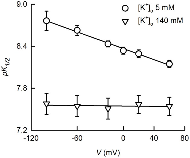
TASK-2 (K2P5.1) is a two-pore domain K(+) channel belonging to the TALK subgroup of the K2P family of proteins. TASK-2 has been shown to be activated by extra- and intracellular alkalinization. Extra- and intracellular pH-sensors reside at arginine 224 and lysine 245 and might affect separate selectivity filter and inner gates respectively. TASK-2 is modulated by changes in cell volume and a regulation by direct G-protein interaction has also been proposed. Activation by extracellular alkalinization has been associated with a role of TASK-2 in kidney proximal tubule bicarbonate reabsorption, whilst intracellular pH-sensitivity might be the mechanism for its participation in central chemosensitive neurons. In addition to these functions TASK-2 has been proposed to play a part in apoptotic volume decrease in kidney cells and in volume regulation of glial cells and T-lymphocytes. TASK-2 is present in chondrocytes of hyaline cartilage, where it is proposed to play a central role in stabilizing the membrane potential. Additional sites of expression are dorsal root ganglion neurons, endocrine and exocrine pancreas and intestinal smooth muscle cells. TASK-2 has been associated with the regulation of proliferation of breast cancer cells and could become target for breast cancer therapeutics. Further work in native tissues and cells together with genetic modification will no doubt reveal the details of TASK-2 functions that we are only starting to suspect.

Keywords: K2P channels; TASK-2 channel; bicarbonate reabsorption; cell volume regulation; central chemoception; chondrocytes.


Figures

Figure 1
Figure 1
TASK-2 position within the K2P phylogeny and structural molecular model. (A) Phylogenetic tree constructed using human K2P channel sequences with the MEGA5 software (www.megasoftware.net) using the maximum likelihood method. The scale bar indicates an evolutionary distance of 0.2 amino acid substitutions per position. Common and International Union of Pharmacology (Goldstein et al., 2005) names are given. (B) Modeling the extracellular ion pathway (EIP) of TASK-2 channel and the position of the R224 pHo sensors. A molecular model for the TASK-2 pore based on the structure of TRAAK (PDB ID 3UM7) is shown with both sensing R224 residues facing the EIP. Shown is a ribbon representation with K+ ions and H2O molecules in the selectivity filter (SF). R224 pHo-sensing residues are shown in stick representation. The EIP is drawn as a solid tunnel connecting the extracellular space and the entrance of the SF. HOLE color code is used: blue, radius >1.15 Å. The illustration is taken at the end of a 10-ns MD run.
Figure 2
Figure 2
K+- and voltage-dependence of TASK-gating by pHo. The dependence of K+ currents upon extracellular pH was studied on HEK-293 cells previously transfected with TASK-2 cDNA. Measurements were done in the whole-cell recording mode of the patch-clamp technique as previously described (Niemeyer et al., 2007). The intracellular solution contained 140 mM K+ whilst extracellular K+ was either 5 or 140 mM. K+ 5 mM bath solution contained 135 mM sodium gluconate, 1 mM potassium gluconate, 4 mM KCl, 2 mM CaCl2, 1 mM MgCl2, 105 mM sucrose, 10 mM HEPES/Tris, pH 7.5. Intracellular, pipette solution contained 8 mM KCl, 132 mM potassium gluconate, 1 mM MgCl2, 10 mM EGTA, 1 mM Na3ATP, 0.1 mM GTP, 10 mM HEPES/Tris, pH7.4. Extracellular pH-dependence curves were generated at various voltages and fits of the Hill equation yielded the pK1/2 values reported which are averages of fitted parameters of the individual experiments. Some of the experiments for the data presented here have been published before (Niemeyer et al., 2010). Use of the Woodhull (1973) model [pK1/2(V) = pK1/2(0 mV)-zδ FV/2.303RT, where pK1/2(V) and K1/2(0 mV) are the −log of the inhibitory dissociation constants at a given voltage V and at 0 mV; δ is the fractional distance across the electric field crossed by H+; and z, R, T, and F have the conventional meaning], on the data at 5 mM K+, resulted in a δ-value of 0.22 and pK1/2(0 mV) of 8.37.
Figure 3
Figure 3
Cartilage as a site of TASK-2 expression revealed by β-galactosidase activity in TASK-2 KO mice. Skeletons from WT (A) and TASK-2 KO (B) mice were prepared by clearing and decalcification followed by staining for X-gal (blue). The KO mouse was generated by a gene-trap approach where the trapping vector encodes a β-galactosidase/neomycin resistance fusion protein (Araki et al., 2009). The enzyme is expressed under the control of the TASK-2 promoter and therefore the β-galactosidase activity reflects sites of endogenous TASK-2 gene expression.
Figure 4
Figure 4
TASK-2 expression in hyaline cartilage from trachea and articular surface of the knee. (A,B) are images of whole trachea stained with X-gal in wild type and heterozygous TASK-2(+/−) mice, respectively. The signal is present in the cartilage rings. In (D) the image demonstrates staining in chondrocytes [(C): wild-type control]. (E,F): images obtained from control and heterozygous TASK-2(+/−) mouse knees. The macroscopic staining in the area of the epiphyseal growth plate observed in WT knee corresponds to cells that are expressing endogenous mammalian ß-gal activity, most likely osteoclasts (Kopp et al., 2007). Bars in (C,D) correspond to 25 μm.

References

    1. Alper S. L. (2010). Familial renal tubular acidosis. J. Nephrol. 23Suppl. 16, S57–S76 - PubMed
    1. Alvarez-Baron C. P., Jonsson P., Thomas C., Dryer S. E., Williams C. (2011). The two-pore domain potassium channel KCNK5: induction by estrogen receptor alpha and role in proliferation of breast cancer cells. Mol. Endocrinol. 25, 1326–1336 10.1210/me.2011-0045 - DOI - PMC - PubMed
    1. Añazco C., Peña-Münzenmayer G., Araya C., Cid L. P., Sepúlveda F. V. (2013). G protein modulation of K2P potassium channel TASK-2: a role of basic residues in the C-terminus domain. Pflugers Arch. [Epub ahead of print]. 10.1007/s00424-00013-1314-0 - DOI - PubMed
    1. Araki M., Araki K., Yamamura K. (2009). International Gene Trap Project: towards gene-driven saturation mutagenesis in mice. Curr. Pharm. Biotechnol. 10, 221–229 10.2174/138920109787315006 - DOI - PubMed
    1. Armstrong C. M. (2003). Voltage-gated K channels. Sci. STKE 2003:re10 10.1126/stke.2003.188.re10 - DOI - PubMed

LinkOut - more resources