. 2010 Oct 31;1(2):e0015.
doi: 10.5041/RMMJ.10015. Print 2010 Oct.

MRI-Guided Electrophysiology Intervention

Affiliations

MRI-Guided Electrophysiology Intervention

Henry R Halperin et al. Rambam Maimonides Med J. .

Erratum in

Abstract

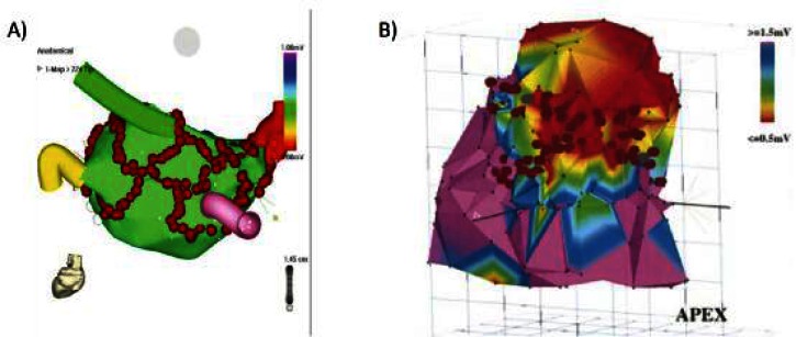
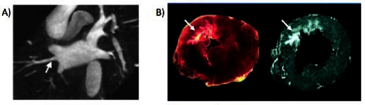
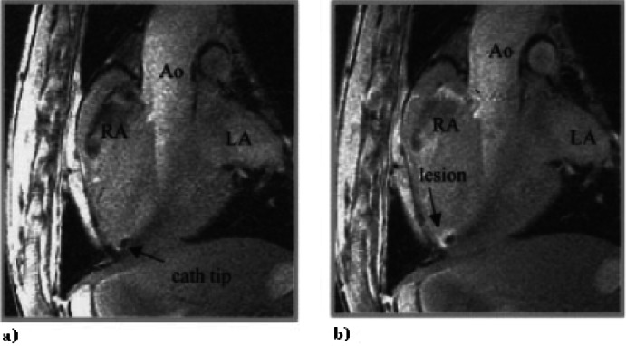
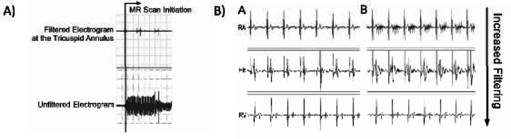
Catheter ablation is a first-line treatment for many cardiac arrhythmias and is generally performed under X-ray fluoroscopy guidance. However, current techniques for ablating complex arrhythmias such as atrial fibrillation and ventricular tachycardia are associated with sub-optimal success rates and prolonged radiation exposure. Pre-procedure 3-D magnetic resonance imaging (MRI) has improved understanding of the anatomic basis of complex arrhythmias and is being used for planning and guidance of ablation procedures. A particular strength of MRI compared to other imaging modalities is the ability to visualize ablation lesions. Post-procedure MRI is now being applied to assess ablation lesion location and permanence with the goal of identifying factors leading to procedure success and failure. In the future, intra-procedure real-time MRI, together with the ability to image complex 3-D arrhythmogenic anatomy and target additional ablation to regions of incomplete lesion formation, may allow for more successful treatment of even complex arrhythmias without exposure to ionizing radiation. Development of clinical grade MRI-compatible electrophysiology devices is required to transition intra-procedure MRI from preclinical studies to more routine use in patients.

Keywords: ablation; arrhythmia; electrophysiology; magnetic resonance imaging.


Figures

Figure 1
Figure 1
Examples of electrospatial mapping guidance of complex arrhythmia ablation. A and B: Electrospatial surface maps generated by point-by-point contact mapping of the endocardial surface. The red circles are markers where ablation energy was delivered. A: Example of atrial fibrillation ablation in which ablations lesions are placed to encircle the pulmonary veins to prevent exit of arrhythmia triggering foci originating from the pulmonary veins. The pulmonary vein locations are marked by the colored “cartoon” tubes. B: Example of scar based ventricular tachycardia ablation in which linear lesions were placed connecting scar (red) to normal tissue (purple) to interrupt the arrhythmia circuit. Figure 1A included with permission from The Journal of Cardiovascular Electrophysiology. (Calkins JCEP 2005; 13:53) Figure 1B included with permission from Circulation (Marchlinski, Circulation 2000; 101:1288).
Figure 2
Figure 2
Examples of arrhythmogenic anatomy depicted by MRI. A: MRI angiogram anatomy of the pulmonary veins. Note that variant pulmonary vein anatomy such as an additional right middle pulmonary vein, indicated by the white arrow, can be clearly seen by MRI. B: The complex structure of myocardial infarction scar, indicated by the white arrows, depicted by pathology on the left and delayed gadolinium enhanced MRI on the right. Figure 2A included with permission from the Journal of Cardiovascular Electrophysiology (Mansour, JCEP 2004; 15:387). Figure 2B included with permission from Circulation (Kim, Circulation 1999; 100:1992).
Figure 3
Figure 3
Example of MRI visualization of an ablation catheter positioned at the right ventricle (RV) apex (a) before and (b) after radiofrequency ablation. Post-ablation images were obtained after peripheral injection of gadolinium contrast. Figure included with permission from Circulation (Lardo, Circulation 2000;102(6):698).
Figure 4
Figure 4
A: Example of the bipolar intracardiac electrograms during scanning before filtering (bottom trace) and after filtering (top trace). B: Example of bipolar intracardiac electrograms at various locations in the heart outside the scanner (left column) and during scanning with increasing levels of filtering (right column). RA = Right atrium, His = His bundle, RV = right ventricular apex.
Figure 5
Figure 5
Example of non-contrast T2-weighted MR imaging of right ventricular epicardial RF ablation lesions with pathologic correlation. Stability of the imaged lesion size is demonstrated from 30 minutes to 12 hours after ablation. Figure included with permission from Heart Rhythm (Dickfeld, HR 2007;4(2):215).
Figure 6
Figure 6
Example of gadolinium-enhanced T1-weighted MR imaging of right ventricular epicardial RF ablation lesions with pathologic correlation. Different lesion enhancement patterns are seen from 1 minute to 2 hours after contrast injection. Figure included with permission from the Journal of the American College of Cardiology (Dickfeld, JACC 2006;47(2):370).
Figure 7
Figure 7
Example of using automatic catheter highlighting and reference image planes to navigate complex 3-D anatomy using real-time MRI. The anatomic location of the catheter position on the image labeled LAX2 is better appreciated when overlaid with long and short axis images of the heart. Figure included with permission from The Journal of Magnetic Resonance Imaging (Guttman, JMRI 2007; 26:1429).

References

    1. Calkins H, Yong P, Miller JM, et al. Catheter ablation of accessory pathways, atrioventricular nodal reentrant tachycardia, and the atrioventricular junction: final results of a prospective, multicenter clinical trial. The Atakr Multicenter Investigators Group. Circulation. 1999;99:262–70. - PubMed
    1. Shah DC, Jais P, Haissaguerre M, et al. Three-dimensional mapping of the common atrial flutter circuit in the right atrium. Circulation. 1997;96:3904–12. - PubMed
    1. Natale A, Newby KH, Pisano E, et al. Prospective randomized comparison of antiarrhythmic therapy versus first-line radiofrequency ablation in patients with atrial flutter. J Am Coll Cardiol. 2000;35:1898–904. doi: 10.1016/S07351097(00)00635-5. - DOI - PubMed
    1. Pappone C, Rosanio S, Oreto G, et al. Circumferential radiofrequency ablation of pulmonary vein ostia: A new anatomic approach for curing atrial fibrillation. Circulation. 2000;102:2619–28. - PubMed
    1. Marchlinski FE, Callans DJ, Gottlieb CD, Zado E. Linear ablation lesions for control of unmappable ventricular tachycardia in patients with ischemic and nonischemic cardiomyopathy. Circulation. 2000;101:1288–96. - PubMed

LinkOut - more resources