. 2011 Jan 31;2(1):e0021.
doi: 10.5041/RMMJ.10021. Print 2011 Jan.

Milestones in the evolution of hepatic surgery

Affiliations

Milestones in the evolution of hepatic surgery

Henri Bismuth et al. Rambam Maimonides Med J. .

Abstract

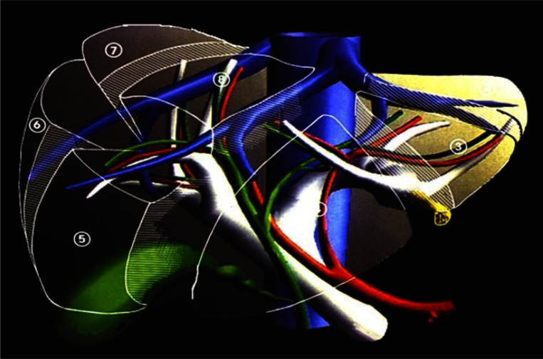
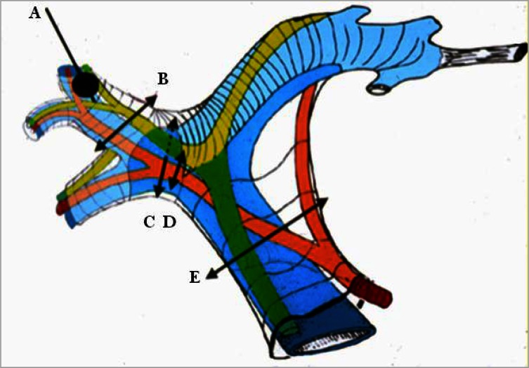
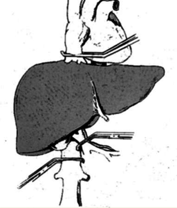
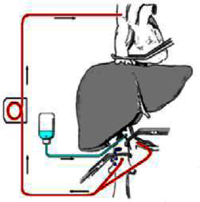
This paper describes the rapid evolution of modern liver surgery, starting in the middle of the twentieth century. Claude Couinaud studied and described the segmental anatomy of the liver, Thomas Starzl performed the first liver transplantations, and Henri Bismuth introduced the concept of anatomical resections. Hepatic surgery has developed significantly since those early days. To date, innovative techniques are applied, using cutting-edge technologies: Intraoperative ultrasound, techniques of vascular exclusion of the liver, new devices for performing homeostasis and dissection, laparoscopy for resections, and new drugs that allow the resection of previously unresectable tumors. The next stage in liver surgery will probably be the implementation of a multidisciplinary holistic approach to the liver-diseased patient that will ensure the best and most efficient treatments in the future.

Keywords: Surgery; liver; transection; transplantation.


Figures

Figure 1
Figure 1
The transparent liver showing the main arteries (in red), portal vein (in white), hepatic veins (in blue), and main parts of the biliary system (in green). Liver segments are numbered, and the round ligament is designated in yellow.
Figure 2
Figure 2
Glissonian pedicle elements: portal vein (in blue), hepatic artery (in red), and the bile ducts (in green). Hepatic inflow occlusion: A) Selective occlusion of segmental portal vein by a balloon introduced under ultrasonographic guidance. The arrows show the different sites of Glissonian clamping. B) Suprahilar clamping of one sector of the right liver; C and D) hilar selective clamping to the right liver; E) total pedicular clamping (Pringle maneuver).
Figure 3
Figure 3
Total vascular exclusion of the liver by clamping the infrahepatic and suprahepatic inferior vena cava and the hepatoduodenal ligament.
Figure 4
Figure 4
Total vascular exclusion of the liver with hypothermia as described by Fortner et al. The liver is excluded (as in Figure 3). Veno-venous bypass of the liver is performed (red lines), and hypothermic solution is infused into the portal vein (in blue).
Figure 5
Figure 5
Total vascular exclusion for complex liver resections. A) The ex-situex-vivo technique described by Pichlmayr et al. B) The ex-situin-vivo technique described by Hannoun et al.

References

    1. Langenbuch C. Ein Fall von Resecktion eines linksseitigen Schnurlappens der Leber. Berl Klin Wochenschr. 1888;25:37. German.
    1. Quattlebaum JK, Quattlebaum JK., Jr Technic of hepatic lobectomy. Ann Surg. 1959;149:648–51. doi: 10.1097/00000658-195905000-00005. - DOI - PMC - PubMed
    1. Fineberg C, Goldburgh WP, Templeton JY., 3rd Right hepatic lobectomy for primary carcinoma of the liver. Ann Surg. 1956;144:881–92. doi: 10.1097/00000658-195611000-00013. - DOI - PMC - PubMed
    1. Lortat-Jacob JL, Robert HG. Well defined technic for right hepatectomy. Presse Med. 1952;60:549–51. French. - PubMed
    1. Couinaud C. Lobes et segments hépatiques. Presse Med. 1954;62:709. French. - PubMed

LinkOut - more resources