Review
. 2013 Aug;9(3):211-9.
doi: 10.2174/1573403x113099990032.

Coarctation of the aorta - the current state of surgical and transcatheter therapies

Affiliations
Review

Coarctation of the aorta - the current state of surgical and transcatheter therapies

Jeffrey E Vergales et al. Curr Cardiol Rev. 2013 Aug.

Abstract

Aortic coarctation represents a distinct anatomic obstruction as blood moves from the ascending to the descending aorta and can present in a range of ages from infancy to adulthood. While it is often an isolated and discrete narrowing, it can also be seen in the more extreme scenario of severe arch hypoplasia as seen in the hypoplastic left heart syndrome or in conjunction with numerous other congenital heart defects. Since the first description of an anatomic surgical repair over sixty years ago, an evolution of both surgical and transcatheter therapies has occurred allowing clinicians to manage and treat this disease with excellent results and low morbidity and mortality. This review focuses on the current state of both transcatheter and surgical therapies, paying special attention to recent data on long-term follow-up of both approaches. Further, current thoughts will be explored about future therapeutic options that attempt to improve upon historical long-term outcomes.
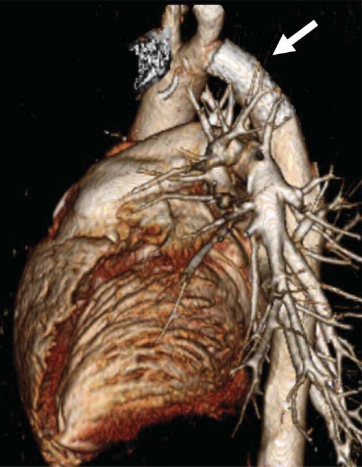

Figures

Fig. (1)
Fig. (1)
Angiogram of aortic coarctation in an infant. The solid black arrow denotes discrete coarctation just distal to the takeoff of the left subclavian artery in what is commonly the juxtaductal position.
Fig. (2)
Fig. (2)
Panel A shows a two-dimensional transthoracic echocardiogram from the suprasternal view demonstrating a discrete narrowing just distal to the take-off of the left subclavian artery. This individual did not have a displaced left subclavian artery but instead had an aberrant right subclavian artery originating near the coarctation (not shown). Note the continuous high velocity color Doppler signal across the hypoplastic coarctation segment (Panel B).
Fig. (3)
Fig. (3)
Magnetic resonance imaging of a 13 year-old female with a history of coarctation repair by end-to-end anastomosis as an infant. Hypertension, with a 20 mmHg extremity gradient seen during follow-up, led to further evaluation which revealed a discrete area of recoarctation (white arrow). She successfully underwent endovascular stenting and is currently doing well.
Fig. (4)
Fig. (4)
Angiogram demonstrating aneurysm development (Panel A, black arrow) in a 13 year-old patient who previously underwent subclavian flap aortoplasty for coarctation of the aorta. Magnetic resonance imaging of a 30 year-old patient in Panel B also demonstrates aneurysm formation (white arrow) after subclavian flap-aortoplasty. Note the absence of the left subclavian artery in both images.
Fig. (5)
Fig. (5)
Recurrent coarctation shown in this 1-year old patient who previously underwent repair by subclavian flap aortoplasty. The white arrow demonstrates the absent left subclavian artery, while the black arrow demonstrates the recurrent coarctation. Balloon angioplasty successfully reduced the gradient across the obstruction from 80 mmHg to 15mmHg. This patient further has a common origin of the left carotid artery and the innominate artery, a common aortic arch variant.
Fig. (6)
Fig. (6)
This patient with a ventricular septal defect and coarctation of the aorta previously underwent ventricular septal defect surgical patch closure and coarctation repair. Recurrence of the coarctation in a long-segment, however, necessitated placement of an interposition graft (black arrow). Recurrent obstruction is seen at the proximal end of the graft (white arrow). Note pacemaker wires related to post-surgical heart block.
Fig. (7)
Fig. (7)
This 1 year-old infant presented with post-operative recurrent coarctation of the aorta (Panel A, black arrow) after patch aortoplasty. A high-pressure angioplasty balloon is inflated across the obstructed segment (Panel B), with improvement angiographically (Panel C) and hemodynamically (pressure gradient reduced from 55 mmHg to 10 mmHg).
Fig. (8)
Fig. (8)
This 8 year-old patient with native coarctation of the aorta, as shown in Panel A, had a 50 mmHg gradient across the narrowed segment (black arrow). He underwent angioplasty with stent placement (Panel B, white arrow), completely eliminating the pressure gradient as well as the anatomic obstruction (Panel C).
Fig. (9)
Fig. (9)
Computed tomography scan with three-dimensional reconstruction in a 22 year-old patient with recurrent coarctation. Note the presence of an endovascular stent in the descending aorta (white arrow) with no proximal aneurysm formation.

References

    1. Kieth J, Rowe R, Vlad P. Heart Disease in Infancy and Childhood. 1958;1:877.
    1. Anderson RH, Lenox CC, Zuberbuhler JR. Morphology of ventricular septal defect associated with coarctation of aorta. Brit Heart J. 1983;50(2):176–81. - PMC - PubMed
    1. Folger GM, Stein PD. Bicuspid aortic valve morphology when associated with coarctation of the aorta. Catheter Cardiovasc Diag. 1984;10(1):17–25. - PubMed
    1. Shone JD, Sellers RD, Anderson RC, Adams P, Lillehei CW, Edwards J. The developmental complex of “parachute mitral valve,” supravalvular ring of left atrium, subaortic stenosis, and coarctation of aorta. Am J Cardiol. 1963;11:714–25. - PubMed
    1. Machii M, Becker A. Nature of coarctation in hypoplastic left heart syndrome. Ann Thorac Surg. 1995;59(6):1491–4. - PubMed