. 2013 Aug;88(8):871-83.
doi: 10.1016/j.mayocp.2013.04.001.

Innate regeneration in the aging heart: healing from within

Affiliations

Innate regeneration in the aging heart: healing from within

Piero Anversa et al. Mayo Clin Proc. 2013 Aug.

Abstract

The concept of the heart as a terminally differentiated organ incapable of replacing damaged myocytes has been at the center of cardiovascular research and therapeutic development for the past 50 years. The progressive decline in myocyte number as a function of age and the formation of scarred tissue after myocardial infarction have been interpreted as irrefutable proofs of the postmitotic characteristic of the heart. However, emerging evidence supports a more dynamic view of the heart in which cell death and renewal are vital components of the remodeling process that governs cardiac homeostasis, aging, and disease. The identification of dividing myocytes in the adult and senescent heart raises the important question concerning the origin of these newly formed cells. In vitro and in vivo findings strongly suggest that replicating myocytes derive from lineage determination of resident primitive cells, supporting the notion that cardiomyogenesis is controlled by activation and differentiation of a stem cell compartment. It is the current view that the myocardium is an organ permissive of tissue regeneration mediated by exogenous and endogenous progenitor cells.

Keywords: CSC; EF; cardiac stem cell; ejection fraction; hCSC; human cardiac stem cell.


Figures

FIGURE 1
FIGURE 1
Heart failure and myocardial regeneration. A, Area of regenerating myocardium in the infarct. Proliferating, small, developing myocytes are positive for the cell-cycle marker MCM5 (white). Myocytes are labeled by cardiac myosin (red) and nuclei by DAPI (blue). Two small myocytes in mitosis are shown in the insets (magnification 300×). B, The cluster of cells included in a rectangle in the middle of an acute infarct is shown at higher magnification in C and D. These cells express c-kit (green, arrows) and at times cardiac myosin (red; D), reflecting undifferentiated cardiac stem cells and early committed progenitors, respectively. MI = myocardial infarction. Scale bars: A and B, 100 µm; C and D, 10 µm.
FIGURE 2
FIGURE 2
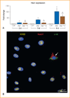
Notch1 up-regulates Nkx2.5 but not GATA4 during early commitment of cardiac stem cells (CSCs). A, Hes1 transcript was measured in CSCs at 2, 5, and 8 days under control conditions (Ctrl) and in the presence of Jagged1 (Jag1) and Jag1 and γ-secretase inhibitor (Jag1 + γ). Hes1 quantity is shown as fold changes vs the corresponding Ctrl. B, After Jag1 stimulation, CSCs are c-kit negative and display nuclear localization of the active fragment of Notch1 (Notch1 intracellular domain [N1ICD], yellow). One CSC continues to express c-kit (green) together with the extracellular domain of the Notch1 receptor (red, arrow). C and D, Jag1-stimulated CSCs express N1ICD (green) together with Nkx2.5 (red). The area included in the square is shown at higher magnification in the inset (magnification: 1000×). E and F, After γ-secretase inhibition, Jag1-stimulated CSCs are negative for N1ICD and Nkx2.5. G, Percentage of CSCs positive for N1ICD and Nkx2.5 at 2, 5, and 8 days. H and I, The expression of GATA4 (yellow) is shown in CSCs stimulated by Jag1 in the (H) absence and (I) presence of γ-secretase inhibitor. J, Percentage of CSCs positive for GATA4 at 5 days. K, Nkx2.5 and GATA4 transcripts were analyzed in CSCs at 5 days. mRNA = messenger RNA. *P<.05 vs Ctrl. **P<.05 vs Jag1. ***Statistically different vs both Ctrl and Jag1. Data are shown as mean ± SD.
FIGURE 2
FIGURE 2
Notch1 up-regulates Nkx2.5 but not GATA4 during early commitment of cardiac stem cells (CSCs). A, Hes1 transcript was measured in CSCs at 2, 5, and 8 days under control conditions (Ctrl) and in the presence of Jagged1 (Jag1) and Jag1 and γ-secretase inhibitor (Jag1 + γ). Hes1 quantity is shown as fold changes vs the corresponding Ctrl. B, After Jag1 stimulation, CSCs are c-kit negative and display nuclear localization of the active fragment of Notch1 (Notch1 intracellular domain [N1ICD], yellow). One CSC continues to express c-kit (green) together with the extracellular domain of the Notch1 receptor (red, arrow). C and D, Jag1-stimulated CSCs express N1ICD (green) together with Nkx2.5 (red). The area included in the square is shown at higher magnification in the inset (magnification: 1000×). E and F, After γ-secretase inhibition, Jag1-stimulated CSCs are negative for N1ICD and Nkx2.5. G, Percentage of CSCs positive for N1ICD and Nkx2.5 at 2, 5, and 8 days. H and I, The expression of GATA4 (yellow) is shown in CSCs stimulated by Jag1 in the (H) absence and (I) presence of γ-secretase inhibitor. J, Percentage of CSCs positive for GATA4 at 5 days. K, Nkx2.5 and GATA4 transcripts were analyzed in CSCs at 5 days. mRNA = messenger RNA. *P<.05 vs Ctrl. **P<.05 vs Jag1. ***Statistically different vs both Ctrl and Jag1. Data are shown as mean ± SD.
FIGURE 3
FIGURE 3
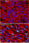
Transit-amplifying myocytes. Small developing myocytes in the infarct are positive for (A) telomerase (magenta) and MCM5 (white) and for (B) MEF2C (yellow) and connexin 43 (green, arrowheads). TERT = telomerase reverse transcriptase. Scale bars, 10 mm.
FIGURE 4
FIGURE 4
Cardiac niches in the human heart. A–C, Cluster of c-kit–positive cardiac stem cells (CSCs) (green). Arrows in A define the areas shown at higher magnification in B and C (magnification 1600×). Gap junctions (connexin 43 [C×43], white; arrowheads) and adherens junctions (N-cadherin [N-cadh], magenta; arrowheads) are illustrated. Connexin 43 and N-cadh are present between CSCs and myocytes (α-sarcomeric actin [SA], red) and fibroblasts (procollagen, light blue; asterisks). Fibronectin, yellow. qdot = quantum dots.
FIGURE 5
FIGURE 5
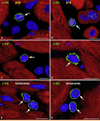
Cardiac stem cell (CSC) senescence. A–F, Two examples each of c-kit–positive CSCs (green) expressing (A and B) p16INK4a (yellow) and (C and D) p53 (magenta) are shown. E and F, Similarly, telomeres in c-kit–positive CSCs are illustrated in 2 examples by small white fluorescence dots. Arrows indicate c-kit–positive CSCs. Scale bars, 10 µm.

Similar articles

Cited by

References

    1. Anversa P, Leri A, Kajstura J. Cardiac regeneration. J Am Coll Cardiol. 2006;47(9):1769–1776. - PubMed
    1. Murry CE, Field LJ, Menasché P. Cell-based cardiac repair: reflections at the 10-year point. Circulation. 2005;112(20):3174–3183. - PubMed
    1. Laflamme MA, Murry CE. Regenerating the heart. Nat Biotechnol. 2005;23(7):845–856. - PubMed
    1. Rubart M, Field LJ. Cardiac regeneration: repopulating the heart. Annu Rev Physiol. 2006;68:29–49. - PubMed
    1. Hansson EM, Lindsay ME, Chien KR. Regeneration next: toward heart stem cell therapeutics. Cell Stem Cell. 2009;5(4):364–377. - PubMed