. 2013 Aug 12;24(2):213-228.
doi: 10.1016/j.ccr.2013.06.014. Epub 2013 Aug 1.

Hexokinase 2 is required for tumor initiation and maintenance and its systemic deletion is therapeutic in mouse models of cancer

Affiliations

Hexokinase 2 is required for tumor initiation and maintenance and its systemic deletion is therapeutic in mouse models of cancer

Krushna C Patra et al. Cancer Cell. .

Erratum in

  • Cancer Cell. 2013 Sep 9;24(3):399

Abstract

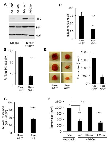
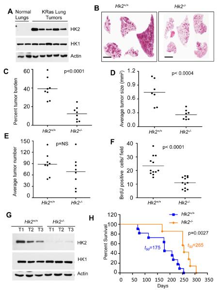
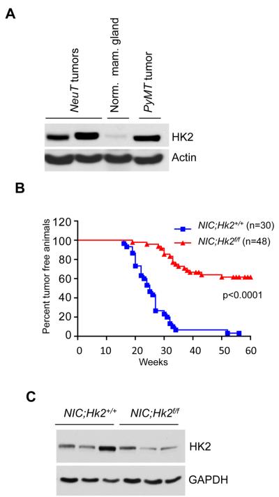
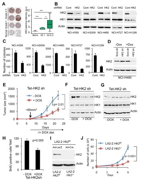
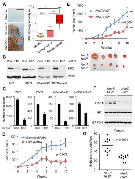
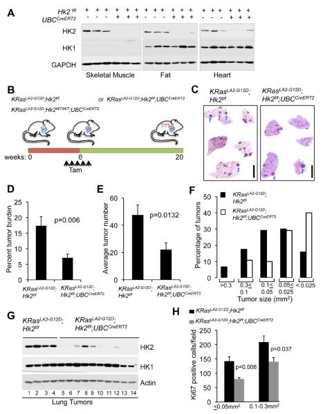
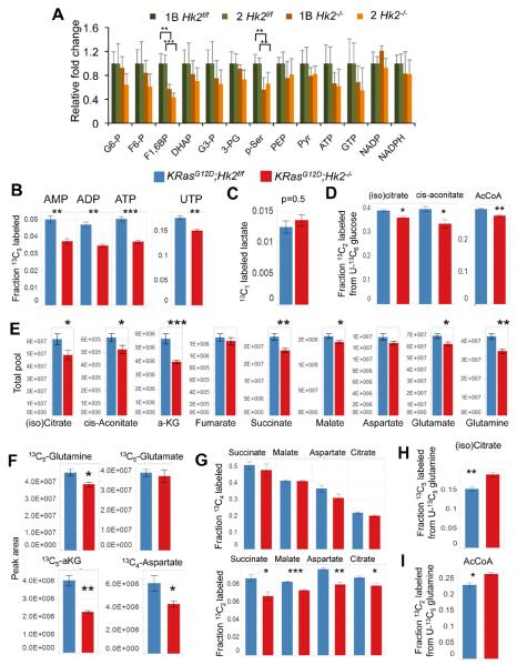
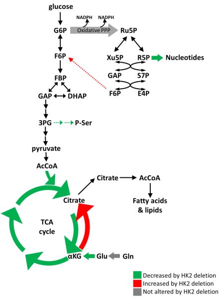
Accelerated glucose metabolism is a common feature of cancer cells. Hexokinases catalyze the first committed step of glucose metabolism. Hexokinase 2 (HK2) is expressed at high level in cancer cells, but only in a limited number of normal adult tissues. Using Hk2 conditional knockout mice, we showed that HK2 is required for tumor initiation and maintenance in mouse models of KRas-driven lung cancer, and ErbB2-driven breast cancer, despite continued HK1 expression. Similarly, HK2 ablation inhibits the neoplastic phenotype of human lung and breast cancer cells in vitro and in vivo. Systemic Hk2 deletion is therapeutic in mice bearing lung tumors without adverse physiological consequences. Hk2 deletion in lung cancer cells suppressed glucose-derived ribonucleotides and impaired glutamine-derived carbon utilization in anaplerosis.


Figures

Figure 1
Figure 1. HK2 is required for oncogenic transformation
A. Immortalized Hk2f/f MEFs following infection with either Ad-Cre or Ad-LacZ, as a control, were established and stably infected with a retrovirus expressing oncogenic HRasV12. Cell extracts were subjected to immunoblotting with anti-HK2 or anti-HK1 antibodies. Control undeleted cells are denoted Hk2f/f and Hk2 deleted cells are denoted Hk2−/−. B. Relative total hexokinase activity in Ras-transformed Hk2f/f and Hk2−/− MEFs. C. Relative glucose consumption in Ras-transformed Hk2f/f and Hk2−/− MEFs. Data represent the percentage of glucose consumed by Ras-transformed Hk2−/− MEFs as compared to Ras-transformed Hk2f/f MEFs after of culturing for 18h, and after adjusting for cell number. D. Average number of colonies generated by Ras transformed Hk2f/f and Hk2−/− MEFs after six weeks growth on soft-agar. E. The effect of Hk2 deletion on tumorigenesis in vivo. Ras-transformed Hk2f/f and Hk2−/− MEFs were injected subcutaneously into athymic nude mice, and tumor size was calculated. Left panel showed representative tumor images of Ras-transformed Hk2f/f and Hk2−/− MEFs (Scale bars: 1 cm). Right panel show the average tumor size formed by Ras-transformed Hk2f/f and Hk2−/− MEFs. (n=4 tumors per each cell type). F. The requirement of HK2 activity for tumorigenesis in vivo. Ras-transformed Hk2−/− MEFs stably expressing WT or kinase-dead HK2, and Ras-transformed Hk2f/f or Hk2−/− control MEFs were subjected to tumorigenesis in vivo as described in (E). Tumor size was calculated when tumor size of control Ras-transformed Hk2f/f MEFs reached approximately 2cm3. (n=4 tumors per each cell type). The data presented in Figs. B–F are expressed as ± SEM where ***, ** and * represent p≤ 0.001, 0.01 and 0.05 respectively. See also Fig. S1.
Figure 2
Figure 2. HK2 is required for the development of oncogenic KRas-driven NSCLC in mice, and its deletion extends the lifespan of these mice
A. Tissue lysates from lungs of control KRasLSL-G12D mice or from tumors in lungs of KRasLSL-G12D mice after exposure to Ad-Cre were subjected to immunoblotting with anti-HK2 or anti-HK1 antibodies. Ad-Cre treated KRasLSL-G12D and KRasLSL-G12D;Hk2f/f mice are denoted as Hk2+/+ and Hk2−/− respectively. B. Sixteen weeks after Cre instillation to induce lung tumors, lung sections were subjected to H&E staining (scale bar 4mm). C. Percentage tumor burden calculated from the H&E stained sections of tumor bearing lungs from KRasLSL-G12D mice (n=8) or KRasLSL-G12D;Hk2f/f mice (n=8). D. Average tumor size per lungs of KRasLSL-G12D (n=7) or KRasLSL-G12D;Hk2f/f (n=8) mice. E. Tumors number calculated from the H&E stained sections of tumor bearing lungs from KRasLSL-G12D (n=8) or KRasLSL-G12D;Hk2f/f (n=8) mice. F. Average number of BrdU positive cells per field as calculated from 40X magnification of tumor sections. At least ten fields of KRasLSL-G12D or KRasLSL-G12D;Hk2f/f lungs from three different mice were used for this analysis. Data in Figs C–F are expressed as the mean ± SEM. G. Immunoblot showing HK2 and HK1 expression in tumors obtained from KRasLSL-G12D or KRasLSL-G12D;Hk2f/f lungs. H. Kaplan-Meier survival curves of lung tumor bearing KRasLSL-G12D or KRasLSL-G12D;Hk2f/f mice after exposure to Ad-Cre at 8 weeks of age (p values and median survival (t=50) for the indicated genotypes were calculated by log-rank test).
Figure 3
Figure 3. HK2 is required for the development of ErbB2/Neu-driven mammary tumors in mice
A. Immunoblot showing HK2 expression in tissue lysates from normal mammary gland or mammary gland tumors isolated from MMTVNeuT and MMTVPyMT mice. B. Kaplan-Meier curves of percentage tumor free survival of MMTVNIC (NIC;Hk2+/+) and MMTVNIC;Hk2f/f (NIC;Hk2f/f) mice. p value was calculated by log-rank test. C. Immunoblot showing HK2 expression in tumor lysates obtained from mice with the indicated genotypes.
Figure 4
Figure 4. HK2 silencing reverses tumorigenesis of NSCLC cells in vitro and in-vivo
A. Left panel: Immunohistochemical staining of HK2 expression in representative human NSCLC patient samples tissue microarray (TMA) correlates with the pathological grade of the disease (scale bar 1mm). Right panel: Quantification of HK2 staining intensity in accordance with the pathological grade. TMA samples analyzed are; 20 non-tumor, 22 grade-1, 44 grade-2, and 14 grade-3. Box plots represent the 25th to 75th percentiles (boxes) with median and the whiskers represent the maximum and the minimum value. B. Immunoblot showing the level of HK2 and HK1 protein expression in a panel of human NSCLC cells stably expressing either shRNA targeting HK2 or LacZ shRNA as control. C. Equal numbers of either control or HK2 knockdown cells were subjected to soft-agar assay, and colonies were counted after three weeks. Data are expressed as the mean of three independent experiments ± SEM (p ≤ 0.002). D. Immunoblot showing HK2 protein level in NCI-H460 cells expressing an inducible control or HK2 shRNA. The cells were exposed to vehicle or 500ng/ml doxycycline (DOX) for 96 hrs prior to analysis. E. NCI-H460 cells (0.75×10⁶) were injected subcutaneously into athymic nude mice. When tumor size reached ~65mm3, mice were fed doxycycline containing diet (n=8) or normal chow diet (n=7), and tumor growth was followed until tumor size reached 1.5–2cm3. F. At the end of the experiment described in E, tumor lysates were subjected to immunoblotting with anti-HK2 antibodies. G. Immunoblot showing HK2 protein levels in tumor lysates harvested one week after exposure to the DOX diet. H. BrdU incorporation in tumors one week after exposure to DOX diet. I. Deletion of Hk2 in NSCLC cell line derived from KRasLA2-G12D;Hk2f/f mice. J. Cell proliferation after Hk2 deletion in the NSCLC cell line derived from KRasLA2-G12D;Hk2f/f mice. Data presented in panels C, E, H and J are expressed as the mean ± SEM. See also Fig. S2.
Figure 5
Figure 5. HK2 silencing reverses tumorigenesis of breast cancer cells in vitro and in vivo
A. Left panel: Representative images of immunohistochemical staining of HK2 in human breast cancer samples TMA, showing low expression in the stroma and benign tumors and high expression in cancer samples (scale bar 0.5mm). Right panel: Quantification of HK2 staining intensity in the stroma, benign tumors and cancer samples (Stroma (n=4, Benign (n=8), Tumor (n=15)). Box plots represent the 25th to 75th percentiles (boxes) with median and the whiskers represent the maximum and the minimum value. B. Immunoblot showing HK2 protein level in a panel of human breast cancer or transformed mammary cells stably expressing either shRNA targeting HK2 or LacZ shRNA as control. C. Equal numbers of control or HK2 knockdown cells were subjected to soft-agar assay and colonies were counted after three weeks. Data is expressed as the mean of three independent experiments ± SEM (p ≤ 0.003). D. Equal numbers of MDA-MB-453 cells stably expressing either HK2 shRNA or control shRNA were injected into the fat pad of the mammary glands of female athymic nude mice. Tumor growth was followed. E. Cells of cell line derived from a mammary tumor of MMTVNeuT;Hk2f/f mice were infected with either Ad-GFP or Ad-Cre-GFP to delete Hk2 and to generate NeuT;Hk2f/f or NeuT;Hk2−/− cells. The cells were then injected orthotopically in female athymic nude mice (6 mice per group) and tumor growth was followed until tumor size reached approximately 2cm3. Bottom panel show representative images of tumors (scale bar 1cm). F. Immunoblot showing HK2 and HK1 protein levels in tumor protein lysates from individual tumors shown in E. G. Quantification of BrdU incorporation in tumor sections derived from the tumors described in E. The average number of BrdU positive cells per field was calculated from (40X magnification) of the tumor tissue sections. At least ten fields from three different tumors per each mouse genotype were used for the quantification. The data in Fig. 5D, 5E and 5G are expressed as the mean ± SEM. See also Fig. S3.
Figure 6
Figure 6. Systemic deletion of Hk2 in the mouse is therapeutic for oncogenic KRas-driven NSCLC
A. Immunoblot showing HK2 and HK1 protein expression in tissues isolated from either Hk2f/f or Hk2f/f;UBCCreERT2 mice two weeks after final tamoxifen injection. B. Schematic illustration of the experiment described in C–H. C. Representative images of H&E staining of whole-mount lung sections prepared from either KRasLA2-G12D;Hk2f/f or KRasLA2-G12D;Hk2f/f;UBCCreERT2 lungs, 12 weeks after injection of tamoxifen at 8 weeks of age. Individual tumors are indicated by arrows (scale bar 4mm). D. Percentage tumor burden as calculated from the H&E sections of tumor bearing lungs from either KRasLA2-G12D;Hk2f/f or KRasLA2-G12D;Hk2f/f;UBCCreERT2 mice at 20 weeks of age and after injection of tamoxifen at 8 weeks of age (n=8 per group). E. Tumor numbers calculated from the H&E stained sections of tumor bearing lungs from either KRasLA2-G12D;Hk2f/f or KRasLA2-G12D;Hk2f/f;UBCCreERT2 mice (n=8 per group). F. Size distribution of tumors developed in the lungs of either KRasLA2-G12D;Hk2f/f or KRasLA2-G12D;Hk2f/f;UBCCreERT2 mice (n=6 per group). G. Immunoblot showing HK2 and HK1 protein levels in tumor lysates extracted at 20 weeks of age, and after tamoxifen injection at 8 weeks of age. Lanes 1–4- tumors were randomly selected for analysis; lanes 5,6 – tumor size < 0.025mm2; lanes 7–9- tumor size 0.3–0.05mm2; lanes 10–14-tumor size 0.05–0.025mm2. H. Quantification of Ki67 staining of the same size groups of tumors derived from either KRasLA2-G12D;Hk2f/f or KRasLA2-G12D;Hk2f/f;UBCCreERT2 mice. Data presented in Figs. D, E and H are expressed as the mean ± SEM. See also Fig. S4.
Figure 7
Figure 7. The effect of Hk2 deletion in KRasG12D NSCLC cells on the intracellular levels of metabolites derived from glucose or glutamine
A. Steady state levels of glycolytic metabolites in LA2-1B and LA2-2 cells in the presence or absence of HK2. Data are presented as fold changes of Hk2f/f versus Hk2−/− (n=5). B. Deletion of Hk2 results in a decreased fraction of 13C5 UTP and 13C5 AMP, ADP and ATP from the PPP upon U-13C6 glucose feeding. C. No significant change between the fraction of labeled lactate containing 13C1, or relative contribution of the oxidative PPP, upon deletion of Hk2. D. Contribution of glucose to the citrate / isocitrate, cis-aconitate and acetyl CoA pools is decreased by Hk2 deletion, as observed by decreased 13C2 species. E. Total pools of TCA cycle metabolites were averaged across all four labeling conditions (U-12C6 Glc; U-13C6 Glc; 1,2-13C2 Glc; U-13C5 Gln. Average of n=16 with SE. F. The 13C5 or 13C4 LC/MS/MS peak areas upon U-13C5 Gln feeding, G. Labeled 13C4 (upper panel) and 13C2 (bottom panel) succinate, malate, aspartate, and citrate derived from glutamine. H. Citrate/isocitrate pools from reductive carboxylation upon Hk2 deletion. I. AcCoA pools from reductive carboxylation upon Hk2 deletion. A–I. Unless otherwise noted, values are an average of n=4 with SE, 2 hours post media switch (p values for A–I are: *p<0.05, **p<0.005, ***p<0.0005). See also Fig. S5.
Figure 8
Figure 8
Schematic illustration depicting metabolic changes induced by Hk2 deletion in KRasG12D-NSCLC cells. Decreased flux is green; increased flux is red; maintained flux is grey. Dashed arrows are predicted to be altered.

References

    1. Cheong H, Lu C, Lindsten T, Thompson CB. Therapeutic targets in cancer cell metabolism and autophagy. Nat Biotechnol. 2012;30:671–678. - PMC - PubMed
    1. DeBerardinis RJ, Mancuso A, Daikhin E, Nissim I, Yudkoff M, Wehrli S, Thompson CB. Beyond aerobic glycolysis: transformed cells can engage in glutamine metabolism that exceeds the requirement for protein and nucleotide synthesis. Proc Natl Acad Sci U S A. 2007;104:19345–19350. - PMC - PubMed
    1. Felig P, Wahren J. Fuel homeostasis in exercise. N Engl J Med. 1975;293:1078–1084. - PubMed
    1. Fueger PT, Heikkinen S, Bracy DP, Malabanan CM, Pencek RR, Laakso M, Wasserman DH. Hexokinase II partial knockout impairs exercise-stimulated glucose uptake in oxidative muscles of mice. Am J Physiol Endocrinol Metab. 2003;285:E958–963. - PubMed
    1. Gaglio D, Metallo CM, Gameiro PA, Hiller K, Danna LS, Balestrieri C, Alberghina L, Stephanopoulos G, Chiaradonna F. Oncogenic K-Ras decouples glucose and glutamine metabolism to support cancer cell growth. Mol Syst Biol. 2011;7:523. - PMC - PubMed

Publication types