. 2013 Oct:63:122-34.
doi: 10.1016/j.yjmcc.2013.07.014. Epub 2013 Aug 1.

Coronary adventitial cells are linked to perivascular cardiac fibrosis via TGFβ1 signaling in the mdx mouse model of Duchenne muscular dystrophy

Affiliations

Coronary adventitial cells are linked to perivascular cardiac fibrosis via TGFβ1 signaling in the mdx mouse model of Duchenne muscular dystrophy

Nicholas Ieronimakis et al. J Mol Cell Cardiol. 2013 Oct.

Abstract

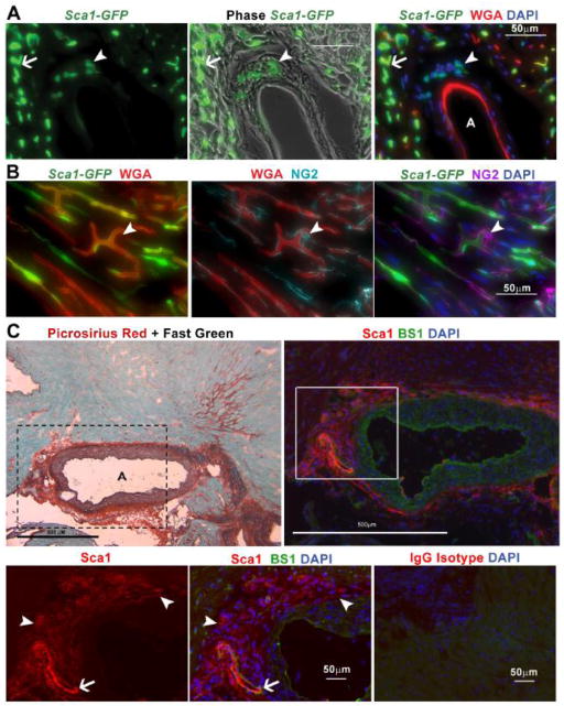
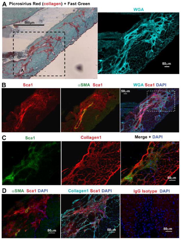
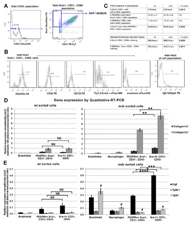
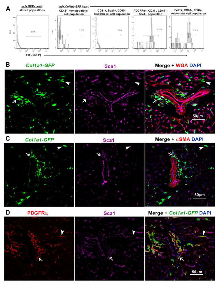
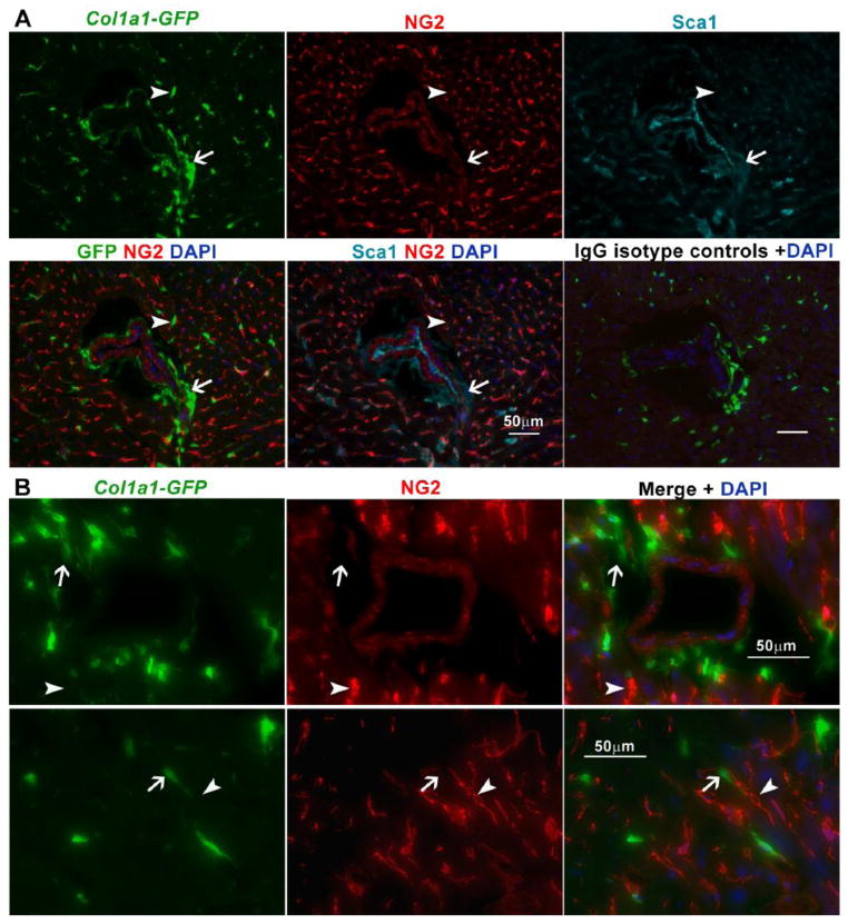
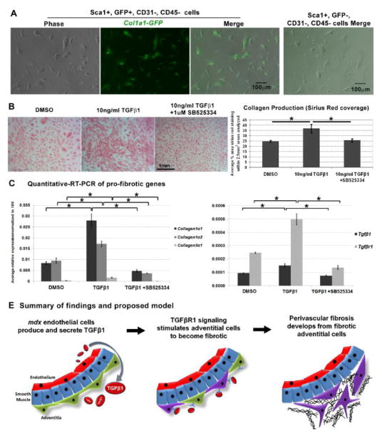
In Duchenne muscular dystrophy (DMD), progressive accumulation of cardiac fibrosis promotes heart failure. While the cellular origins of fibrosis in DMD hearts remain enigmatic, fibrotic tissue conspicuously forms near the coronary adventitia. Therefore, we sought to characterize the role of coronary adventitial cells in the formation of perivascular fibrosis. Utilizing the mdx model of DMD, we have identified a population of Sca1+, PDGFRα+, CD31-, and CD45- coronary adventitial cells responsible for perivascular fibrosis. Histopathology of dystrophic hearts revealed that Sca1+ cells extend from the adventitia and occupy regions of perivascular fibrosis. The number of Sca1+ adventitial cells increased two-fold in fibrotic mdx hearts vs. age matched wild-type hearts. Moreover, relative to Sca1-, PDGFRα+, CD31-, and CD45- cells and endothelial cells, Sca1+ adventitial cells FACS-sorted from mdx hearts expressed the highest level of Collagen1α1 and 3α1, Connective tissue growth factor, and Tgfβr1 transcripts. Surprisingly, mdx endothelial cells expressed the greatest level of the Tgfβ1 ligand. Utilizing Collagen1α1-GFP reporter mice, we confirmed that the majority of Sca1+ adventitial cells expressed type I collagen, an abundant component of cardiac fibrosis, in both wt (71%±4.1) and mdx (77%±3.5) hearts. In contrast, GFP+ interstitial fibroblasts were PDGFRα+ but negative for Sca1. Treatment of cultured Collagen1α1-GFP+ adventitial cells with TGFβ1 resulted in increased collagen synthesis, whereas pharmacological inhibition of TGFβR1 signaling reduced the fibrotic response. Therefore, perivascular cardiac fibrosis by coronary adventitial cells may be mediated by TGFβ1 signaling. Our results implicate coronary endothelial cells in mediating cardiac fibrosis via transmural TGFβ signaling, and suggest that the coronary adventitia is a promising target for developing novel anti-fibrotic therapies.

Keywords: Adventitia; Fibrosis; Muscular dystrophy; Perivascular; Sca1; TGFβ1; Type I collagen.


Conflict of interest statement

Disclosure Statement

The authors concede no conflicts of interest in the publication of this manuscript.

Figures

Fig. 1
Fig. 1
Sca1+ cells, distinct from endothelial cells and pericytes, reside in the coronary adventitia. A. Histological analysis of hearts from 11 month old Sca1-GFP animals injected intravenously with WGA, reveals coronary adventitial cells are Sca1+ (arrowhead) and distinct from GFP+, IV injected-WGA+ endothelial cells (arrow) B. Staining for NG2 indicates pericytes (arrowhead) are negative for Sca1-GFP but cover GFP+, IV injected-WGA+ vascular endothelial cells. C. Staining with picrosirius red and fast green or Sca1 and BS1 in sections from a 22 month old mdx heart, reveals that collagen deposition surrounding the coronary adventitia is occupied by Sca1+, BS1− cells. A higher magnification (boxed area represented in bottom row) photo highlights adventitial Sca1+, BS1− cells (arrowhead) and Sca1+, BS1+ endothelial cells (arrow). The bottom right panel shows a serial section of the same mdx heart shown in C, stained in parallel with an IgG isotype antibody control. Coronary arteries are denoted with an A.
Fig. 2
Fig. 2
Sca1+ cells extend from the adventitia and occupy regions of perivascular fibrosis. A. Picrosirius red-fast green staining reveals perivascular fibrosis in 22 month old mdx hearts extending from coronary arteries. The boxed area depicts a serial section of the same region stained with WGA, which highlights the perivascular fibrosis depicted in by picrosirius red staining. The A denotes a coronary artery. B. Sca1+ cells are observed extending from the adventitia of α-smooth muscle actin+ (αSMA) coronary arteries, to occupy the corresponding fibrotic area that stained brightly positive with WGA (same staining shown in the right panel of A). C. Staining for type I collagen in serial sections from the same heart highlights the occupation of this fibrotic area (boxed region in the merge of B) by Sca1+ cells. D. A higher magnification photo of the same area shown in C, depicts Sca1 with αSMA and collagen1 staining’s respectively. The far right panel depicts a section stained in parallel with an IgG isotype antibody to exclude the presence of non-specific staining.
Fig. 3
Fig. 3
Sca1+ adventitial cells express markers and genes associated with fibrosis. A. Representative flow cytometry gating used for selecting the Sca1+, CD31−, CD45− adventitial population from cell suspensions isolated from wt and mdx hearts. B. analysis of mdx cells using a panel of antibodies shows the Sca1+, CD31−, CD45− population is predominantly PDGFRα + and CD34+, but CD133− and Thy1.2−. Unstained and IgG isoptype controls were used for gating analysis. C. Analysis of heart cells from 1 year old wt (n=4) vs. mdx (n=4) males, revealed that the proportion and absolute number of Sca1+ adventitial cells doubles in mdx hearts. In contrast, the number of PDGFRα+, Sca1−, CD31−, CD45− cells decline, indicating that Sca1+ cells become the predominant PDGFRα+ cells in mdx hearts. D and E. Quantitative-RT-PCR analysis of freshly sorted cells (from1 year old males, n=4 wt and n=4 mdx hearts) reveals that in comparison to endothelial cells (Sca1+, CD31+, CD45-) and macrophages (CD45+, F4/80+), the PDGFRα+, Sca1−, CD31−, CD45− population and Sca1+, CD31−, CD45− adventitial cells both express genes indicative of fibrosis. Although not significantly (NS) different in wt, expression of these genes was significantly elevated in mdx adventitial cells. Surprisingly, endothelial cells from mdx hearts expressed the highest levels of the Tgfβ1 ligand. Each sample was normalized to its respective expression of 18S. * P<0.05, **P<0.005, ***P<0.0005. # denotes P<0.005 between endothelial cells vs. remaining populations. Error bars represent SEM, P values were derived from Student’s t-test.
Fig. 4
Fig. 4
The majority of Sca1+ adventitial cells express Collagen1α1-GFP and are distinguishable from Sca1− interstitial fibroblasts. A. FACS-analysis of mdx:Col1α1-GFP hearts (n=3 GFP+ and n=1 GFP− control, 4 month old males) reveals that the majority of Sca1+, CD31−, CD45− adventitial cells are GFP+. In turn, the highest proportion of GFP+ cells was observed in the Sca1+ adventitial cells vs. other populations analyzed. B. Histological analysis of an aged matched mdx:Col1α1-GFP heart, confirms the presence of Sca1+, GFP+ cells (arrow) in the adventitia of coronary arteries. In contrast, Sca1−, GFP+ interstitial cells (arrowhead) were visible near WGA+, Sca1+ endothelial cells. C. αSMA staining highlights the anatomical location of Sca1+, GFP+ (arrow) adventitial cells vs. Sca1−, GFP+ (arrowhead) interstitial fibroblasts. D. Staining for PDGFRα confirms the presence of PDGFRα+, GFP+ interstitial fibroblasts (arrowhead), distinguishable from Sca1+ adventitial cells (arrow).
Fig. 5
Fig. 5
Collagen1α1-GFP+ cells are adventitial cells and fibroblasts distinct from pericytes. A. Staining for the pericyte marker NG2 indicates that GFP+ cells in mdx:Col1α1-GFP hearts are distinct from pericytes. As shown, Sca1+, GFP+ adventitial cells (arrow) and Sca1−, GFP+ interstitial cells (arrow) were both negative for NG2. The bottom right panel represent mdx:Col1α1-GFP heart tissue stained in parallel with IgG isotype controls for both Sca1 and NG2 antibodies. B. Higher magnification of a separate coronary vessel (top row) and interstitial space (bottom row), highlight the presence of NG2+, GFP− pericytes (arrowhead) vs. GFP+ cells (arrow).
Fig. 6
Fig. 6
Sca1+ adventitial cells maintain expression of Collagen1α1-GFP and become fibrotic in response to TGFβ1 stimulation. A. GFP+, Sca1+, CD31−, CD45− cells FACS-sorted from 4 month old wt Col1α1-GFP hearts maintained expression of GFP in culture. In contrast, GFP-, Sca1+, CD31−, CD45− cells sorted in parallel never expressed Collagen1α1-GFP in culture, right photograph. B. GFP+, Sca1+ sorted cells were treated with the TGFβ1 ligand (10ng/ml) to assess their fibrotic response. In parallel, cells were treated with the vehicle (DMSO) and in conjunction with TGFβ1, an inhibitor of TGFβR1 signaling (SB525334). Each experimental condition was conducted with four replicates (n=4 replicates per condition). Following 4 days of treatment, cells were fixed and stained for collagen with picrosirius red representative photographs that were used for analysis and quantification of collagen production (picrosirius red coverage) with each treatment. Quantification reveals that cells treated with TGFβ1 produced more collagen vs. vehicle controls and cells exposed to 1μM of SB525334. C. In a duplicate experiment, cells were collected for quantitative-RT-PCR analysis of pro-fibrotic genes (n=4 replicates per condition). Analogous to the results in B, cells treated with 10ng/ml TGFβ1 expressed significantly higher levels of Collagen1α1, 1α2 and 3α1 as compared to cells treated with DMSO and 10ng/ml TGFβ1 +1μM SB525334. Error bars represent SEM, *P<0.005 derived from Student’s t-test. D. Model summarizing our results and mechanism of TGFβ1 mediated perivascular fibrosis.

References

    1. Deconinck N, Dan B. Pathophysiology of duchenne muscular dystrophy: current hypotheses. Pediatric neurology. 2007;36:1–7. - PubMed
    1. Culligan KG, Mackey AJ, Finn DM, Maguire PB, Ohlendieck K. Role of dystrophin isoforms and associated proteins in muscular dystrophy (review) International journal of molecular medicine. 1998;2:639–48. - PubMed
    1. Sultan A, Fayaz M. Prevalence of cardiomyopathy in Duchenne and Becker’s muscular dystrophy. J Ayub Med Coll Abbottabad. 2008;20:7–13. - PubMed
    1. Spurney CF. Cardiomyopathy of Duchenne muscular dystrophy: current understanding and future directions. Muscle & nerve. 2011;44:8–19. - PubMed
    1. Eagle M, Baudouin SV, Chandler C, Giddings DR, Bullock R, Bushby K. Survival in Duchenne muscular dystrophy: improvements in life expectancy since 1967 and the impact of home nocturnal ventilation. Neuromuscul Disord. 2002;12:926–9. - PubMed

Publication types

MeSH terms