Review
. 2013 Nov;1830(11):5204-10.
doi: 10.1016/j.bbagen.2013.07.025. Epub 2013 Aug 2.

The glucose signaling network in yeast

Affiliations
Review

The glucose signaling network in yeast

Jeong-Ho Kim et al. Biochim Biophys Acta. 2013 Nov.

Abstract

Background: Most cells possess a sophisticated mechanism for sensing glucose and responding to it appropriately. Glucose sensing and signaling in the budding yeast Saccharomyces cerevisiae represent an important paradigm for understanding how extracellular signals lead to changes in the gene expression program in eukaryotes.

Scope of review: This review focuses on the yeast glucose sensing and signaling pathways that operate in a highly regulated and cooperative manner to bring about glucose-induction of HXT gene expression.

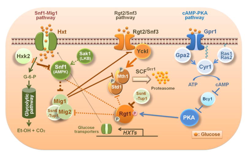
Major conclusions: The yeast cells possess a family of glucose transporters (HXTs), with different kinetic properties. They employ three major glucose signaling pathways-Rgt2/Snf3, AMPK, and cAMP-PKA-to express only those transporters best suited for the amounts of glucose available. We discuss the current understanding of how these pathways are integrated into a regulatory network to ensure efficient uptake and utilization of glucose.

General significance: Elucidating the role of multiple glucose signals and pathways involved in glucose uptake and metabolism in yeast may reveal the molecular basis of glucose homeostasis in humans, especially under pathological conditions, such as hyperglycemia in diabetics and the elevated rate of glycolysis observed in many solid tumors.

Keywords: Cancer; Glucose signaling pathways; Glucose transporters; Glucose uptake and metabolism; Yeast.


Figures

Fig. 1
Fig. 1
Mth1 is required for the interaction of Rgt1 with Ssn6-Tup1 that leads to repression of HXT gene expression; indeed, its inactivation is critical for glucose uptake and metabolism. A) Rgt1 recruits Ssn6-Tup1 in an Mth1-dependent manner and brings about repression of its target genes, such as HXT and HXK2 genes. Std1 is a paralog of Mth1 but has little effect on regulating Rgt1 function. Glucose-induction of HXT gene expression is achieved by a two-step process: (1) Mth1 and Std1 are degraded by the ubiquitin-proteasome pathway, rendering the PKA phosphorylation sites in Rgt1 available for phosphorylation; (2) Rgt1 phosphorylation by PKA induces its dissociation from Ssn6-Tup1 and consequently from HXT promoters. B) Time-lapse observation of Mth1 degradation in high-glucose medium (4%). Mth1 and Std1 are ubiquitinated by the SCFGrr1 ubiquitin-ligase, and the ensuing ubiquitination of Mth1 and Std1 targets them to the proteasome for degradation. The figure was adapted from [45].
Fig. 2
Fig. 2
The two glucose responsive repressors Rgt1 and Mig1 are regulated in a similar manner. A) Rgt1 recruits Ssn6-Tup1 in the absence of glucose; however, it is hyperphosphorylated by PKA in the presence of high levels of glucose and dissociated from Ssn6-Tup1, resulting in the induction of expression of genes involved in glucose uptake and metabolism. B) Ssn6-Tup1 interacts with only unphosphorylated Mig1 in high levels of glucose and mediates the repression of genes involved in glucose oxidation and carbon catabolite repression. Snf1-dependent phosphorylation of Mig1 in glucose-limited conditions abolishes interaction with Ssn6-Tup1 [62].
Fig. 3
Fig. 3
Schematic diagram of the crosstalk between glucose signaling pathways in yeast. Yck I (Yck1 and Yck2) phosphorylates Mth1 and Std1 upon activation by glucose-bound Rgt2 and Snf3 glucose sensors. Phosphorylated Mth1 and Std1 are ubiquitinated by the SCFGrr1 complex and degraded by the proteasome. The PKA phosphorylation sites in the amino terminal region of Rgt1 are exposed and available for phosphorylation when Mth1 is degraded. Phosphorylated Rgt1 is dissociated form Ssn6-Tup1 and subsequently from DNA, leading to derepression of Rgt1 target genes, such as the HXT and HXK2 genes. The Rgt2/Snf3 pathway regulates itself through glucose-induction of STD1 gene expression. Consequently, the STD1 gene is expressed at the same time that the Std1 protein is degraded in response to glucose [13]. By contrast, glucose stimulates Mth1 degradation but also represses Mth1 expression via Mig1 and Mig2. Glucose uptake is required for the generation of the glucose repression signal that leads to inactivation of the Snf1 kinase [18]. Expression of the MIG2 gene is induced by glucose via the Rgt2/Snf3 pathway. Glucose-repression of SNF3 gene expression by Mig1 reflects the probable function of Snf3 as a high affinity glucose sensor, representing another important feature of the interaction between the glucose induction and repression pathways.

References

    1. Ozcan S, Johnston M. Function and regulation of yeast hexose transporters. Microbiol Mol Biol Rev. 1999;63:554–569. - PMC - PubMed
    1. Rolland F, Winderickx J, Thevelein JM. Glucose-sensing mechanisms in eukaryotic cells. Trends Biochem Sci. 2001;26:310–317. - PubMed
    1. Santangelo GM. Glucose signaling in Saccharomyces cerevisiae. Microbiol Mol Biol Rev. 2006;70:253–282. - PMC - PubMed
    1. Busti S, Coccetti P, Alberghina L, Vanoni M. Glucose signaling-mediated coordination of cell growth and cell cycle in Saccharomyces cerevisiae. Sensors. 2010;10:6195–6240. - PMC - PubMed
    1. Towle HC. Glucose as a regulator of eukaryotic gene transcription. Trends Endocrinol Metab. 2005;16:489–494. - PubMed

Publication types

MeSH terms

Substances