Review
. 2014 Feb;20(1):71-81.
doi: 10.1177/1073858413498306. Epub 2013 Aug 2.

Seeing the unseen: the hidden world of slow axonal transport

Affiliations
Review

Seeing the unseen: the hidden world of slow axonal transport

Subhojit Roy. Neuroscientist. 2014 Feb.

Abstract

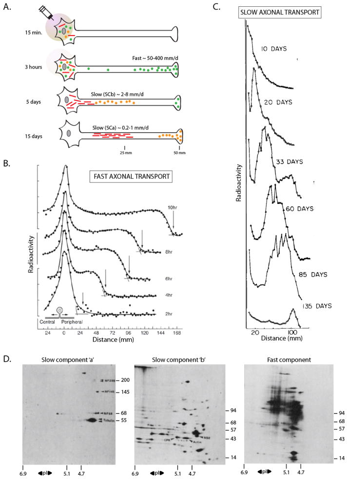
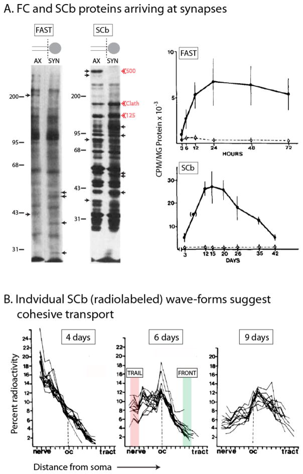
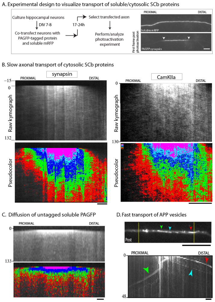
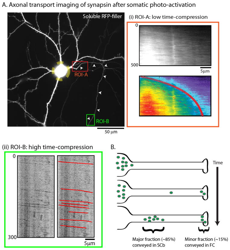
Axonal transport is the lifeline of axons and synapses. After synthesis in neuronal cell bodies, proteins are conveyed into axons in two distinct rate classes-fast and slow axonal transport. Whereas fast transport delivers vesicular cargoes, slow transport carries cytoskeletal and cytosolic (or soluble) proteins that have critical roles in neuronal structure and function. Although significant progress has been made in dissecting the molecular mechanisms of fast vesicle transport, mechanisms of slow axonal transport are less clear. Why is this so? Historically, conceptual advances in the axonal transport field have paralleled innovations in imaging the movement, and slow-transport cargoes are not as readily seen as motile vesicles. However, new ways of visualizing slow transport have reenergized the field, leading to fundamental insights that have changed our views on axonal transport, motor regulation, and intracellular trafficking in general. This review first summarizes classic studies that characterized axonal transport, and then discusses recent technical and conceptual advances in slow axonal transport that have provided insights into some long-standing mysteries.

Keywords: cargo complexes; cytosolic proteins; diffusion; slow axonal transport; soluble proteins; transport packets.


Conflict of interest statement

Conflict of interest: none

Figures

Figure 1
Figure 1. The pulse-chase radiolabeling paradigm to study axonal transport
(A) Radiolabeled amino acids injected in the vicinity of neuronal cell-bodies of an adult animal are incorporated by newly synthesized proteins, and then transported into axons and distal synapses by endogenous processes. The movement of these proteins is then inferred by analyzing sequential axonal segments over incremental time-periods (for specifics of methods, see figure 2 of Roy et al., 2005). After labeling, a population of proteins (green circles) is rapidly conveyed into axons at rates of 50–400 mm/day (‘fast component’, vesicular cargoes). A second pool enters the axons at velocities that are several orders of magnitude lower at 0.2–8 mm/day (‘slow component’). The slow component can be further resolved into two largely distinct ‘peaks’ composed of cytosolic/soluble cargoes (‘Slow Component-b’ or SCb – orange circles) or the major cytoskeletal cargoes (‘Slow Component-a’ or SCa – red bars). (B) Kinetics of fast axonal transport in cat sensory axons accessed by pulse-chase radiolabeling. Note the rapid movement of the radiolabeled wave-front along the peripheral axon over 10 hours (~ 4.5 mm/s). Also note the broad plateau behind the advancing ‘front’ suggesting deposition of cargoes (vesicles) during transit. (C) Kinetics of slow axonal transport in rat motor neurons accessed by pulse-chase radiolabeling. Note the extremely slow movement of the slow component wave-front (~ 100 mm in over 100 days). (D) 2-D PAGE analysis of the three rate components. Transported radiolabeled proteins from mouse or guinea pig optic axons were separated by mass/charge, and analyzed by autoradiography. Some individual protein ‘spots’ are identified by arrowheads on the gels – neurofilaments (NF) and tubulin (SCa, left); creatine phospho-kinase (CPK), actin and non-specific enolase (NSE, SCb, middle). Note the unique overall composition of the three rate-classes. Isoelectric points are on the x-axis and molecular weights are on the y-axis. Figure (B) adapted from Ochs et al., 1981; figure (C) adapted from Hoffman and Lasek, 1975; figure (D) adapted from Brady and Lasek, 1982 – all with permission.
Figure 2
Figure 2. A detailed look at the overall kinetics of cytosolic/soluble cargoes moving in SCb
(A) After somatic pulse-chase radiolabeling, terminal axon (“AX”) or synaptosomal (“SYN’) preparations from guinea pig retinal ganglion cells were analyzed at various time-points to document the ingress of labeled proteins into distal axons (left lanes) and synapses (right lanes). Small arrows point to proteins that are selectively enriched in axons or synapses. Red arrowheads highlight clathrin and two other major unidentified SCb proteins (125 and 500 kDa). In bottom panel, note that the total pool of transported SCb proteins is about three-fold larger than proteins conveyed in the fast component (bottom panel). Also note the decay in radiolabeled SCb proteins after entry into synapses, suggesting turnover and/or retrograde transport at synapses. Figure adapted from Garner and Mahler, 1987, with permission. (B) Overlaid radiolabeled “wave-profiles” of 20 SCb proteins at 4, 6 and 9 days after somatic radiolabeling. Note the striking overlap between different SCb wave-profiles suggesting association with a common “carrier”. Also note the maintenance of the overall coherence of the “fronts” (shaded green) and the “peaks”, even after several days of transit; and also the divergence in the radiolabeled “trails” (shaded pink) suggesting that the deposition of individual moving SCb proteins along the axon was variable. Figures adapted from Garner and Lasek, 1982, with permission.
Figure 3
Figure 3. Photoactivation paradigm to visualize the slow axonal transport of cytosolic SCb protein populations
(A) Cultured neurons are co-transfected with a PAGFP-tagged protein of interest and untagged mRFP (to identify transfected axons). A discrete axonal ROI (~ 20 μm) is photoactivated, and the dispersion of photoactivated molecules is visualized over time (examples of images with PAGFP:synapsin are shown). (B) Greyscale (above) and pseudo-colored (below) kymographs from two PAGFP-tagged SCb proteins synapsin and CamKIIa, imaged using the paradigm above (distance/time in kymographs is on the x/y axis respectively). Note the anterogradely-biased plume of fluorescence. (C) Photoactivation of untagged PAGFP leads to a rapid and unbiased diffusion of fluorescence as expected; different from SCb proteins. (D) Photoactivation of APP – a vesicle-associated fast-component protein – results in the stochastic bidirectional departure of individual vesicles; also different from SCb proteins (colored arrowheads mark the same vesicles in image/kymograph). Scale bar = 5 μm. Figure adapted from Scott et al., 2011 and Tang et al., 2012, with permission.
Figure 4
Figure 4. A fraction of SCb proteins are conveyed in the fast component
(A) To more closely simulate the radiolabeling paradigm, cultured neurons were transfected with GFP:synapsin and soluble mRFP (shown); the neuronal soma was photoactivated (yellow dashed ROI); and the egress of photoactivated molecules into the emergent axon was evaluated over time. While the bulk of synapsin molecules moved slowly into the proximal axon with kinetics expected for slow axonal transport (ROI-A – red box), rapidly-moving particles of synapsin were also seen when the distal axon was imaged after somatic photoactivation (ROI-B – green box). (B) The “dynamic recruitment” model for SCb transport. After synthesis in the soma, cytosolic molecules intermittently and probabilistically associate with “carriers” moving in fast axonal transport. As such, some molecules remain associated with these carriers for long periods, giving rise to a small population (~ 10–15%) that is rapidly transported to axons and synapses. However the majority of cytosolic molecules are slowly conveyed with kinetics resembling slow axonal transport. An implication of this model is that common transport “carriers” are responsible for conveying both fast component and SCb proteins. Figure adapted from Scott et al., 2011, with permission.

References

    1. Allen RD, Metuzals J, Tasaki I, Brady ST, Gilbert SP. Fast axonal transport in squid giant axon. Science. 1982;218:1127–1129. - PubMed
    1. Baas PW, Brown A. Slow axonal transport: the polymer transport model. Trends Cell Biol. 1997;7:380–384. - PubMed
    1. Baitinger C, Willard M. Axonal transport of synapsin I-like proteins in rabbit retinal ganglion cells. J Neurosci. 1987;7:3723–3735. - PMC - PubMed
    1. Black MM, Lasek RJ. Axonal transport of actin: slow component b is the principal source of actin for the axon. Brain Res. 1979;171:401–13. - PubMed
    1. Black MM, Chestnut MH, Pleasure IT, Keen JH. Stable clathrin: uncoating protein (hsc70) complexes in intact neurons and their axonal transport. J Neurosci. 1991;11:1163–1172. - PMC - PubMed

Publication types

MeSH terms