. 2013 Nov 12:252:302-19.
doi: 10.1016/j.neuroscience.2013.07.051. Epub 2013 Jul 31.

Hydrogen sulfide attenuates neurodegeneration and neurovascular dysfunction induced by intracerebral-administered homocysteine in mice

Affiliations

Hydrogen sulfide attenuates neurodegeneration and neurovascular dysfunction induced by intracerebral-administered homocysteine in mice

P K Kamat et al. Neuroscience. .

Abstract

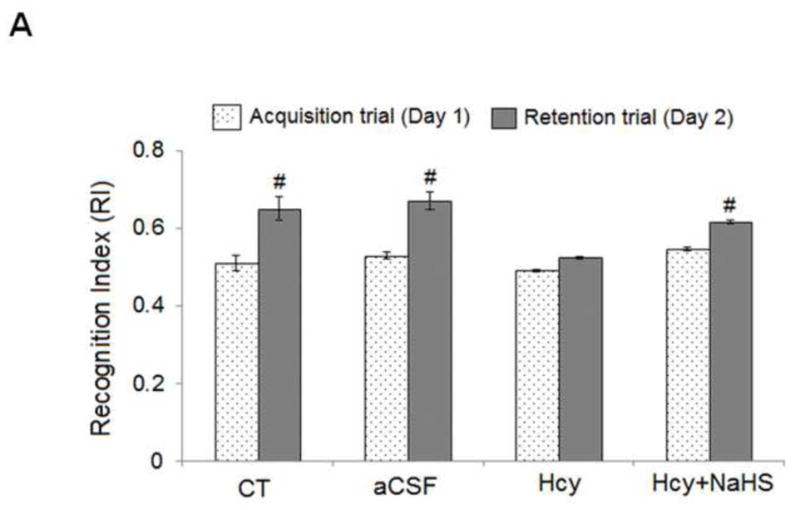
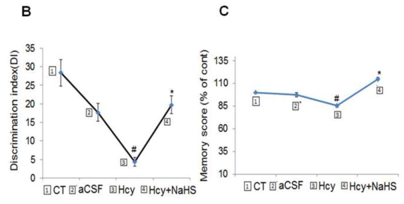
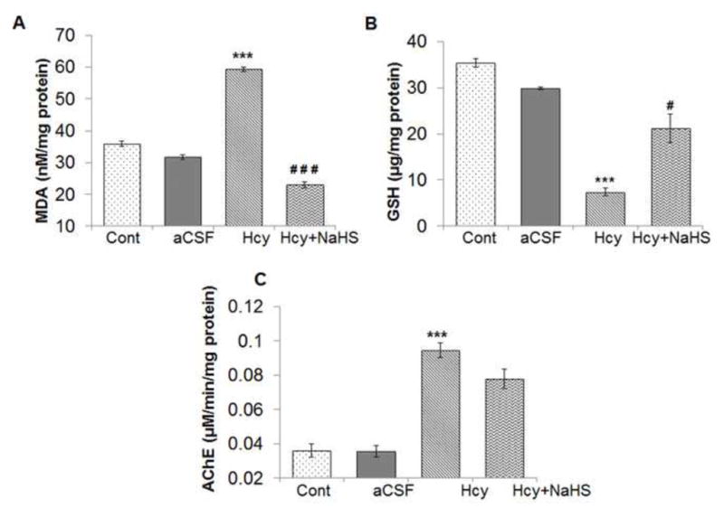
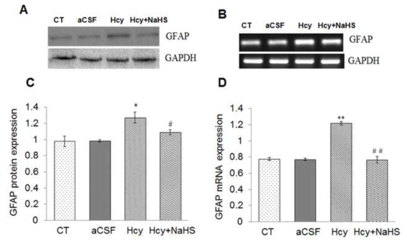
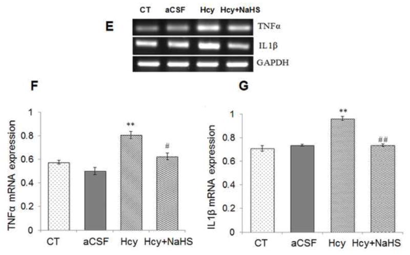
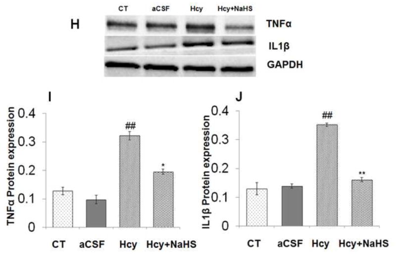
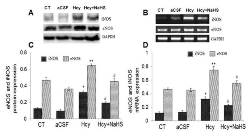
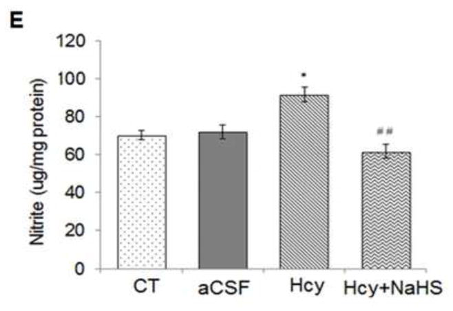
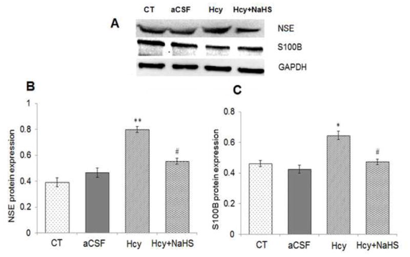
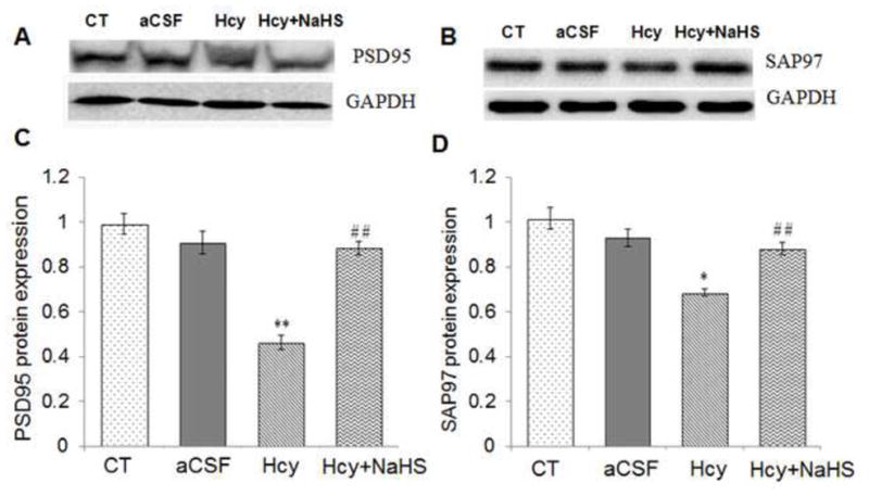
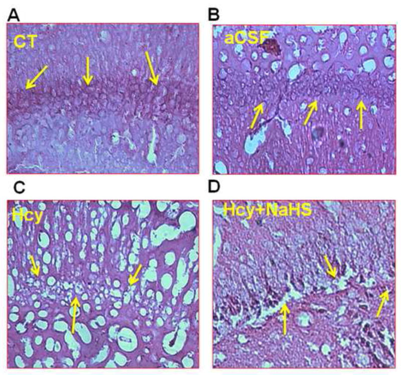
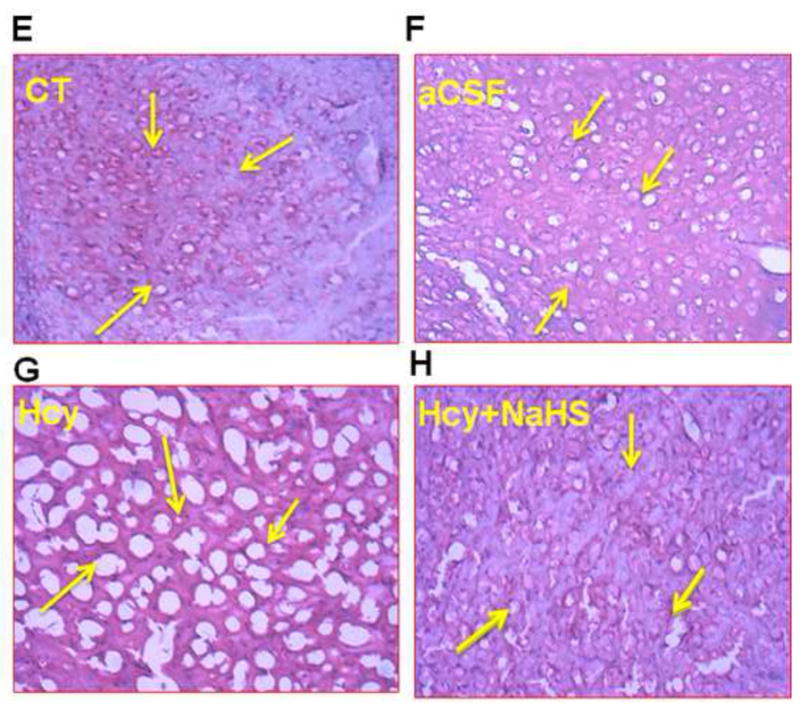
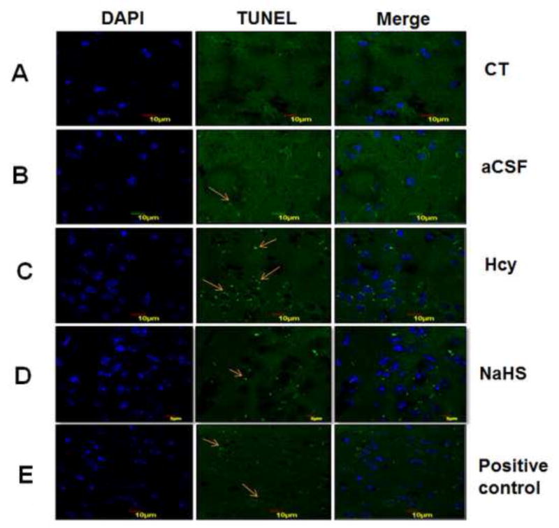
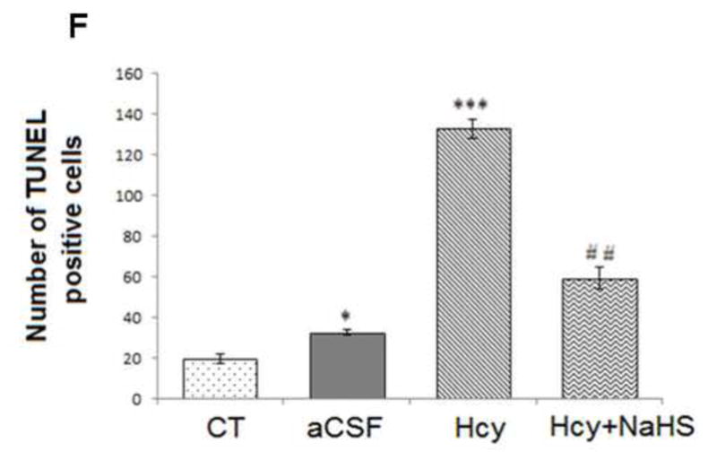
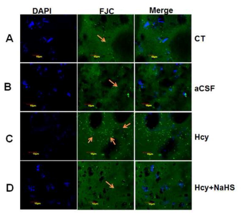
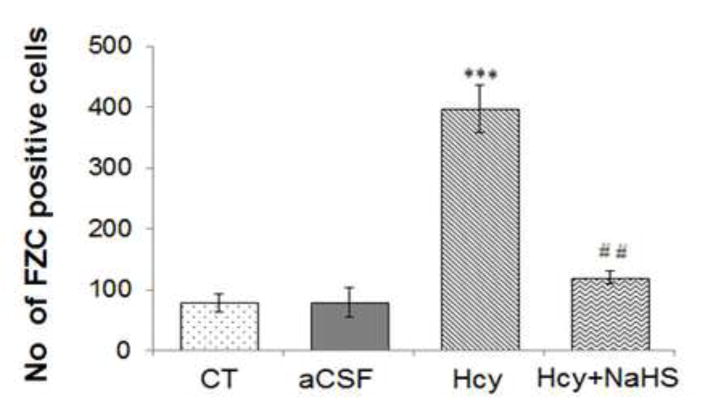
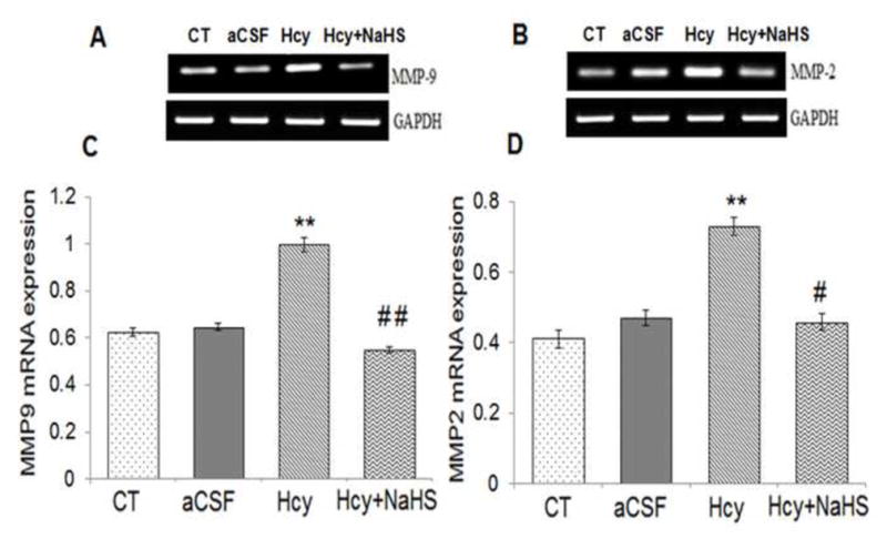
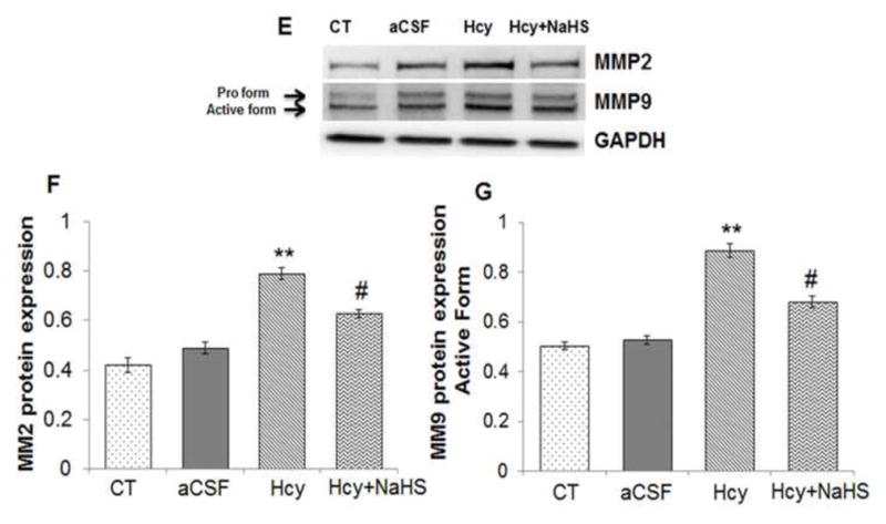
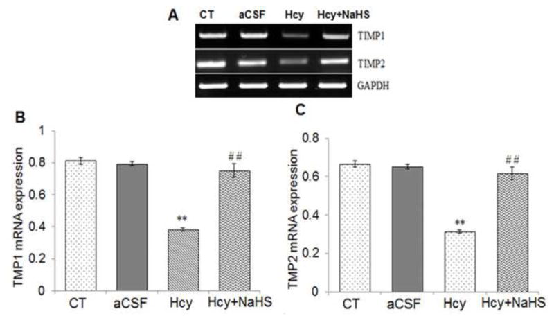
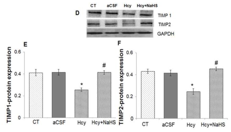
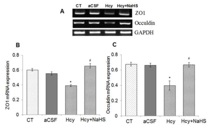
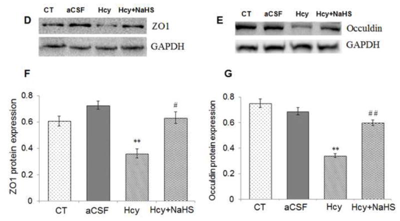
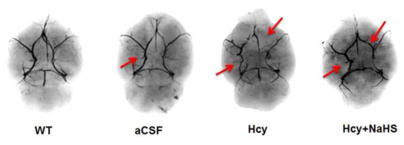
High levels of homocysteine (Hcy), known as hyperhomocysteinemia are associated with neurovascular diseases. H2S, a metabolite of Hcy, has potent anti-oxidant and anti-inflammatory activities; however, the effect of H2S has not been explored in Hcy (IC)-induced neurodegeneration and neurovascular dysfunction in mice. Therefore, the present study was designed to explore the neuroprotective role of H2S on Hcy-induced neurodegeneration and neurovascular dysfunction. To test this hypothesis we employed wild-type (WT) males ages 8-10 weeks, WT+artificial cerebrospinal fluid (aCSF), WT+Hcy (0.5 μmol/μl) intracerebral injection (IC, one time only prior to NaHS treatment), WT+Hcy+NaHS (sodium hydrogen sulfide, precursor of H2S, 30 μmol/kg, body weight). NaHS was injected i.p. once daily for the period of 7 days after the Hcy (IC) injection. Hcy treatment significantly increased malondialdehyde, nitrite level, acetylcholinestrase activity, tumor necrosis factor-alpha, interleukin-1 beta, glial fibrillary acidic protein, inducible nitric oxide synthase, endothelial nitric oxide synthase and decreased glutathione level indicating oxidative-nitrosative stress and neuroinflammation as compared to control and aCSF-treated groups. Further, increased expression of neuron-specific enolase, S100B and decreased expression of (post-synaptic density-95, synaptosome-associated protein-97) synaptic protein indicated neurodegeneration. Brain sections of Hcy-treated mice showed damage in the cortical area and periventricular cells. Terminal deoxynucleotidyl transferase-mediated, dUTP nick-end labeling-positive cells and Fluro Jade-C staining indicated apoptosis and neurodegeneration. The increased expression of matrix metalloproteinase (MMP) MMP9, MMP2 and decreased expression of tissue inhibitor of metalloproteinase (TIMP) TIMP-1, TIMP-2, tight junction proteins (zonula occulden 1) in Hcy-treated group indicate neurovascular remodeling. Interestingly, NaHS treatment significantly attenuated Hcy-induced oxidative stress, memory deficit, neurodegeneration, neuroinflammation and cerebrovascular remodeling. The results indicate that H2S is effective in providing protection against neurodegeneration and neurovascular dysfunction.

Keywords: 5,5′-dithiobis-(2-nitrobenzoic acid); AChE; AD; Alzheimer’s diseases; BBB; DI; DTNB; ECM; EDTA; FJC; Fluro Jade-C; GFAP; GSH; H(2)S; HE; HRP; Hcy; Hematoxylin and Eosin; IC; IL; MDA; MMP; NSE; PBS; PD; PMSF; PSD95; PVDF; Parkinson’s disease; RI; RIPA; RT-PCR; SAP97; TBS-T; TCA; TIMP; TJPs; TNF; TUNEL; Tris-buffered saline with Tween 20; WT; ZO; aCSF; acetylcholinesterase; artificial cerebrospinal fluid; blood–brain barrier; cerebrovascular dysfunction; discrimination index; eNOS; endothelial nitric oxide synthase; ethylenediaminetetraacetic acid; extracellular matrix; glial fibrillary acidic protein; glutathione; homocysteine; horseradish peroxidase; iNOS; inducible nitric oxide synthase; interleukin; intracerebral; malondialdehyde; matrix metalloproteinases; neurodegeneration; neuroinflammation; neuron-specific enolase; phenylmethylsulfonyl fluoride; phosphate-buffered saline; polyvinylidene difluoride; post-synaptic density-95; radio immunoprecipitation assay; recognition index; reverse transcription polymerase chain reaction; synaptosome associated protein-97; terminal deoxynucleotidyl transferase-mediated, dUTP nick-end labeling; tight junction proteins; tissue inhibitor of metalloproteinases; trichloroacetic acid; tumor necrosis factor; wild type; zonula occuldens.


Figures

Fig. 1
Fig. 1. Hcy (IC) induced memory impairment in mice
Mice were subjected to novel object recognition test. Results were expressed as, recognition index (Fig. 1A), discrimination index (Fig. 1B) and mean memory score (Fig. 1C). Data were analyzed by one-way ANOVA followed by Tukey’s test for multiple comparisons.
Fig. 2
Fig. 2. Effect of NaHS on Hcy-induced alterations in malondialdehyde, intracellular reduced glutathione (GSH) and AChE levels
(A) MDA: [F(3, 16)=1.23; P< 0.001] (B) GSH: [F(3, 16)=2.2; P< 0.005] and (C) AChE: [F(3, 16)=1.2; P< 0.01] denotes Hcy significantly increased oxidative stress. A significant protection was observed with NaHS treatment. Data represents mean ±SE from n = 5 per group; ***P< 0.0001 vs control group, #P< 0.05, ###P< 0.0001 vs to Hcy treated group.
Fig. 3
Fig. 3. Effect of NaHS on Hcy induced change in GFAP, TNFα and IL1β
(A) Western blot analyses of GFAP protein expression; [F(3, 16)=.93: P< 0.005] (B) RT-PCR analysis GFAP mRNA expression: [F(3, 16)=1.22; P< 0.01] in WT, WT+aCSF, WT+Hcy and Hcy-treated WT+NaHS mouse brain. The GAPDH was using as a loading control (C& D) Densitometry analysis of GFAP protein/mRNA expressions as represented in the bar diagram. (E) Western blot analyses of TNFα protein expression: [F(3, 16)=1.93; P< 0.01] and IL1β: [F(3, 16)=.73; P< 0.01]. (F) RT-PCR analysis TNFα: [F(3, 16)=.22; P< 0.005] and IL1β mRNA expression: [F(3, 16)=.72; P< 0.001]. The GAPDH was using as a loading control (G& H) Densitometry analysis of TNFα and IL1β protein/mRNA expressions as represented in the bar diagram. Data represents mean ±SE from n = 5 per group. *P< 0.05, **P< 0.005 vs Control group and #P< 0.05, ##P< 0.001 vs Hcy treated group.
Fig. 4
Fig. 4. Effect of NaHS on nitric oxide bioavailability
(A) Western blots analyses of eNOS: [F(3, 16)=1.23; P< 0.01] and iNOS protein: [F(3, 16)=1.5; P< 0.01] (B) RT-PCR analysis of eNOS: [F(3, 16)=2.21; P< 0.005] and iNOS mRNA expression: [F(3, 16)=1.13; P< 0.01] in WT, WT+aCSF, WT+Hcy and Hcy-treated WT+NaHS mouse brain. The GAPDH was using as a loading control (C&D) Densitometry analysis of eNOS and iNOS protein/mRNA expressions as represented in the bar diagram. (E) Nitrite: [F(3, 16)=2.23; P< 0.05]. Data represents mean ±SE from n = 5 per group. *P< 0.05, **P< 0.005 vs Control group and #P< 0.05, ##P< 0.001 vs Hcy treated group
Fig. 5
Fig. 5. Effect of NaHS on neuronal damage markers
(A) Western blots analyses of S100 calcium-binding protein B (S100B): [F(3, 16)=.81; P< 0.01] and neuron-specific enolase (NSE): [F(3, 16)=.33: P< 0.01] in mouse brain. The GAPDH was using as a loading control (B& C) Densitometry analysis of S100B and NSE protein expressions as represented in the bar diagram. Data represents mean ±SE from n = 5 per group. *P< 0.05, **P< 0.005 vs Control group and #P< 0.05, ##P< 0.001 vs Hcy treated group.
Fig. 6
Fig. 6. Effect of NaHS on synaptic proteins
(A) Western blots analyses of PSD95: [F(3, 16)=.61; P< 0.01] (B) SAP97protein expression: [F(3, 16)=.83; P< 0.05] in different treated groups. The GAPDH was using as a loading control (B& C) Densitometry analysis of PSD95 and SAP97 protein expressions as represented in the bar diagram. Data represents mean ±SE from n = 5 per group. *P< 0.05, **P< 0.005 vs Control group and #P< 0.05, ##P< 0.001 vs Hcy treated group.
Fig. 7
Fig. 7. Hematoxylin and eosin (H&E) staining in frozen brain sections in WT, WT+aCSF, WT+Hcy and Hcy-treated WT+NaHS groups
(A) Representative picture of control hippocampus region (B) aCSF did not reveal difference in cell numbers in the neurons of hippocampus region (C) Death of the neurons of hippocampus region revealed degeneration in the hippocampus of Hcy-treated mice (D) Less degeneration is observed in hippocampus region of NaHS treated group. While in periventricular cortex (E) control (F) aCSF did not reveal difference in cortical cells while (G) Hcy administration showed remarkable degeneration (sponginess of the cell) of periventricular cortical neurons (H) NaHS treatment showed less periventricular cortex neuronal degeneration than Hcy treated mice as indicated by arrows (sponginess, condensed nucleus) at 60x magnification. Note: Arrow is showing periventricular cortex neuronal degeneration area.
Fig. 8
Fig. 8. Tunnel assay for apoptosis in WT, WT+aCSF, WT+Hcy and Hcy-treated WT+NaHS groups
Apoptotic cells are seen as green fluorescent dots (scale bar- 10 μm). No difference observed in apoptotic cells of control (A) and CSF (B) group. Apoptotic cell death was increased in Hcy treated brain (C). NaHS treament decreased the apoptotic cell (D). Positive control also represented in this image (E). Bar graph representing a significance of difference between different treatment groups (F). Data represents mean ±SE from n = 5 per group; *P< 0.05, **P< 0.005 vs Control group and #P< 0.05, ##P< 0.001 vs Hcy treated group. Note: Arrow is showing apoptotic cells.
Fig. 9
Fig. 9. Representative Photomicrographs of fluro Jade-C (FJC) staining
(A) Control (B) aCSF did not reveal difference in FJC positive cells (C) A significant increase in FJC positive cell indicates degeneration of neuronal cells in Hcy-treated mice. A significant less numbers of FJC positive cells are observed in NaHS (D) treated mice indicates neuro-protective effect of H2S. Fig. E represents the bar diagram of FJC positive cell (expressed in arbitrary unit). ***P< 0.0001 vs Control group and ##P< 0.005, vs Hcy treated group. Note: Arrow is showing degenerating neurons.
Fig. 10
Fig. 10. Effect of NaHS on MMPs
(A) RT-PCR analysis of MMP-9 mRNA expression: [F(3, 16)=1.29; P< 0.01] (B) RT-PCR analysis of MMP-2 mRNA expression [F(3, 16)=1.33; P< 0.001] in WT, WT+aCSF, WT+Hcy and Hcy-treated WT+NaHS mouse brain. The GAPDH was using as loading control (C&D) Densitometry analysis of MMP-9 and MMP-2 mRNA expressions as represented in the bar diagram. Data represents mean ±SE from n = 5 per group. **P< 0.005 vs Control group and #P< 0.01, vs Hcy treated group. (E) Western blot analysis of MMP-2: [F(3, 16)=1.1; P< 0.001] and MMP-9: [F(3, 16)=.76; P< 0.001] protein expression in different treated groups. The GAPDH was using as loading control (F&G) Densitometry analysis of MMP-2/MMP-9 protein expressions as represented in the bar diagram. Data represents mean ±SE from n = 5 per group. **P< 0.005 vs Control group and #P< 0.01, vs Hcy treated group.
Fig. 11
Fig. 11. Effect of NaHS on TIMPs
(A) RT-PCR analysis of TIMP-1: [F(3, 16)=1.45; P< 0.005] and TIMP-2 mRNA expression: [F(3, 16)=.63; P< 0.01] in WT, WT+aCSF, WT+Hcy and Hcy-treated WT+NaHS mouse brain. The GAPDH was using as loading control (B&C) Densitometry analysis of TIMP-1/TIMP-2 mRNA expressions as represented in the bar diagram. Data represents mean ±SE from n = 5 per group. **P< 0.005 vs Control group and ##P< 0.001 vs Hcy treated group. (D) Western blot analysis of TIMP-1: [F(3, 16)=.34; P< 0.001] and TIMP-2: [F(3, 16)=.55; P< 0.05] protein expression in mouse brain. The GAPDH was using as loading control (E & F) Densitometry analysis of TIMP-1/TIMP-2 protein expressions as represented in the bar diagram. Data represents mean ±SE from n = 5 per group. **P< 0.005 vs Control group and #P< 0.01, ##P< 0.001 vs Hcy treated group.
Fig. 12
Fig. 12. Effect of NaHS on Hcy-induced alterations in tight junction proteins
(A) RT-PCR analysis of ZO-1: [F(3, 16)=.97; P< 0.001] and Occuldin: [F(3, 16)=1.23; P< 0.01] mRNA expression in different treatment groups. The GAPDH was using as loading control (B&C) Densitometry analysis of ZO-1 and Occuldin mRNA expressions as represented in the bar graph (D, E) Western blot analysis of ZO-1: [F(3, 16)=.59; P< 0.001] and Occuldin: [F(3, 16)=.23; P< 0.001] protein expression in WT, WT+aCSF, WT+Hcy and Hcy-treated WT+NaHS mouse brain. Densitometry analysis of ZO-1 and Occuldin protein expressions as represented in the bar diagram (F &G). Data represents mean ±SE from n = 4 per group. **P< 0.005 vs Control group, #P< 0.01, # # P< 0.005 vs Hcy treated group.
Fig. 13
Fig. 13
Brain angiography Study in mice brain showed the loss of major vessel in Hcy treated mice brain which was recovered by NaHS treatment. Note: Arrow showing major blood vessel.

References

    1. Abbott NJ, Patabendige AA, Dolman DE, Yusof SR, Begley DJ. Structure and function of the blood-brain barrier. Neurobiol Dis. 2010;37:13–25. - PubMed
    1. Abbott NJ, Rönnbäck L, Hansson E. Astrocyte-endothelial interactions at the blood-brain barrier. Nat Rev Neurosci. 2006;7:41–53. - PubMed
    1. Alvarez B, Ruiz C, Chacon P, Alvarez-Sabin J, Matas M. Serum values of metalloproteinase-2 and metalloproteinase-9 as related to unstable plaque and inflammatory cells in patients with greater than 70% carotid artery stenosis. J Vasc Surg. 2004;40:469–475. - PubMed
    1. Asahi M, Asahi K, Jung JC, del Zoppo GJ, Fini ME, Lo EH. Role for matrix metalloproteinase 9 after focal cerebral ischemia: effects of gene knockout and enzyme inhibition with BB-94. J Cereb Blood Flow Metab. 2000;20:1681–1689. - PubMed
    1. Ataie A, Sabetkasaei M, Haghparast A, Moghaddam AH, Ataee R, Moghaddam SN. Curcumin exerts neuroprotective effects against homocysteine intracerebroventricular injection-induced cognitive impairment and oxidative stress in rat brain. J Med Food. 2010;13:821–826. - PubMed

Publication types

MeSH terms