Review
. 2013 Jul 31;14(8):15910-30.
doi: 10.3390/ijms140815910.

Magnetic iron oxide nanoparticles for multimodal imaging and therapy of cancer

Affiliations
Review

Magnetic iron oxide nanoparticles for multimodal imaging and therapy of cancer

Reju Thomas et al. Int J Mol Sci. .

Abstract

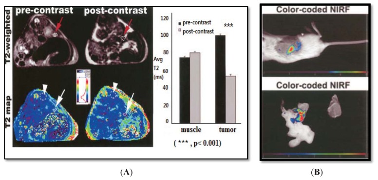
Superparamagnetic iron oxide nanoparticles (SPION) have emerged as an MRI contrast agent for tumor imaging due to their efficacy and safety. Their utility has been proven in clinical applications with a series of marketed SPION-based contrast agents. Extensive research has been performed to study various strategies that could improve SPION by tailoring the surface chemistry and by applying additional therapeutic functionality. Research into the dual-modal contrast uses of SPION has developed because these applications can save time and effort by reducing the number of imaging sessions. In addition to multimodal strategies, efforts have been made to develop multifunctional nanoparticles that carry both diagnostic and therapeutic cargos specifically for cancer. This review provides an overview of recent advances in multimodality imaging agents and focuses on iron oxide based nanoparticles and their theranostic applications for cancer. Furthermore, we discuss the physiochemical properties and compare different synthesis methods of SPION for the development of multimodal contrast agents.


Figures

Figure 1
Figure 1
Concept of multimodal contrast agent based on SPION.
Figure 2
Figure 2
(A) T2 weighted images. The images are before and after the injection of CLIO-EPPT in human pancreatic adenocarcinoma. The T2 relaxation rate is 46.5% (arrow), whereas muscle (arrowhead) is not affected; (B) Color-coded map of NOD/SCID mice bearing orthotopically implanted human pancreatic adenocarcinoma obtained 24 h after I.V. injection of CLIO-EPPT that shows high-intensity fluorescence. Reprinted with permission from [45].
Figure 3
Figure 3
Schematic illustration (a) shows hybrid nanoparticles before and after coating with amphiphilic poly (DMA-r-mPEGMA-r -MA), also shown in TEM images (b) and (c), respectively. CT (d) and MRI (e) images in a hepatoma model at various time points. A white arrow indicates the site of the tumor. Reprinted with permission from [49].
Figure 4
Figure 4
In vivo PET/MRI imaging studies with [64Cu (dtcbp) 2]–Endorem in a mouse. (A) and (B) show signal decreases in popliteal lymph nodes before and after injection indicated by solid arrows; (C) refers to NanoPET/CT images, showing uptake in popliteal (represented by solid arrows) and iliac (represented by hollow arrows) lymph nodes; (D) A whole body NanoPET–CT image of the mouse. Reprinted with permission from [53].
Figure 5
Figure 5
T2-weighted axial MRI for A431K5 tumors. (A), (B), and (C) show pre-injection and 24 and 72 h post-injection MR images. The inset represents autoradiographic images of 20 μM tumor sections; (D) ROI intensity histograms for T2-weighted axial gradient-echo MR images of A431K5 tumors, which show apparent shifts in a 24-h image towards low intensity. Reprinted with permission from [4].
Figure 6
Figure 6
Different approaches adopted in developing a SPION-based theranostic agent.
Figure 7
Figure 7
(a) T2-weighted MR images taken at 0 h and 4.5 h after injection of Dox@TCL-SPION at the LLC tumor on the right back of the mouse; (b) Optical fluorescence images of major organs and allograft tumors: 1 liver; 2 lung; 3 spleen; 4 tumor; 5 heart; and 6 kidney. Images were taken after intravenous injection of Dox@TCL-SPION (equivalent to 4 mg of Dox) (above) and free Dox (4 mg) into tumor-bearing mice (below) (n = 3). The mice were euthanized after 1 h and 12 h. Reprinted with permission from [63].

References

    1. Na H.B., Song I.C., Hyeon T. Inorganic nanoparticles for mri contrast agents. Adv. Mater. 2009;21:2133–2148.
    1. Willmann J.K., van Bruggen N., Dinkelborg L.M., Gambhir S.S. Molecular imaging in drug development. Nat. Rev. Drug Discov. 2008;7:591–607. - PubMed
    1. Kirui D.K., Khalidov I., Wang Y., Batt C.A. Targeted near-ir hybrid magnetic nanoparticles for in vivo cancer therapy and imaging. Nanomed. Nanotechnol. Biol. Med. 2012;9:702–711. - PubMed
    1. Misri R., Meier D., Yung A.C., Kozlowski P., Hafeli U.O. Development and evaluation of a dual-modality (mri/spect) molecular imaging bioprobe. Nanomed. Nanotechnol. Biol. Med. 2012;8:1007–1016. - PubMed
    1. Torres Martin de Rosales R., Tavare R., Glaria A., Varma G., Protti A., Blower P.J. ((9)(9)m)tc-bisphosphonate-iron oxide nanoparticle conjugates for dual-modality biomedical imaging. Bioconjugate Chem. 2011;22:455–465. - PMC - PubMed

Publication types

MeSH terms