. 2013 Jul 31;14(8):15959-76.
doi: 10.3390/ijms140815959.

Effect of δ-opioid receptor activation on BDNF-TrkB vs. TNF-α in the mouse cortex exposed to prolonged hypoxia

Affiliations

Effect of δ-opioid receptor activation on BDNF-TrkB vs. TNF-α in the mouse cortex exposed to prolonged hypoxia

Xuesong Tian et al. Int J Mol Sci. .

Abstract

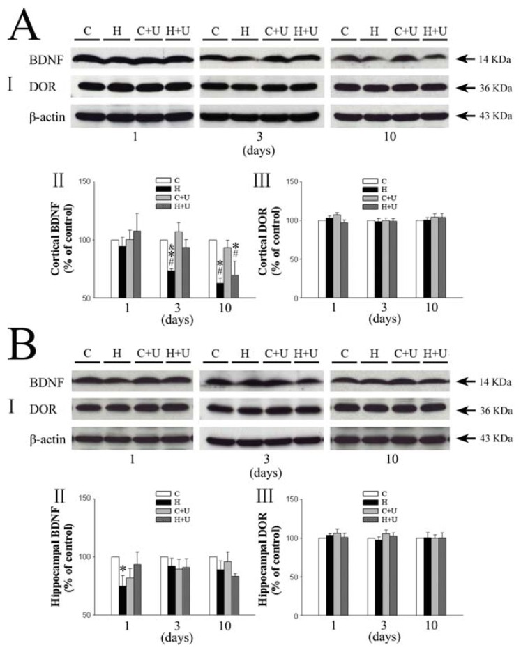
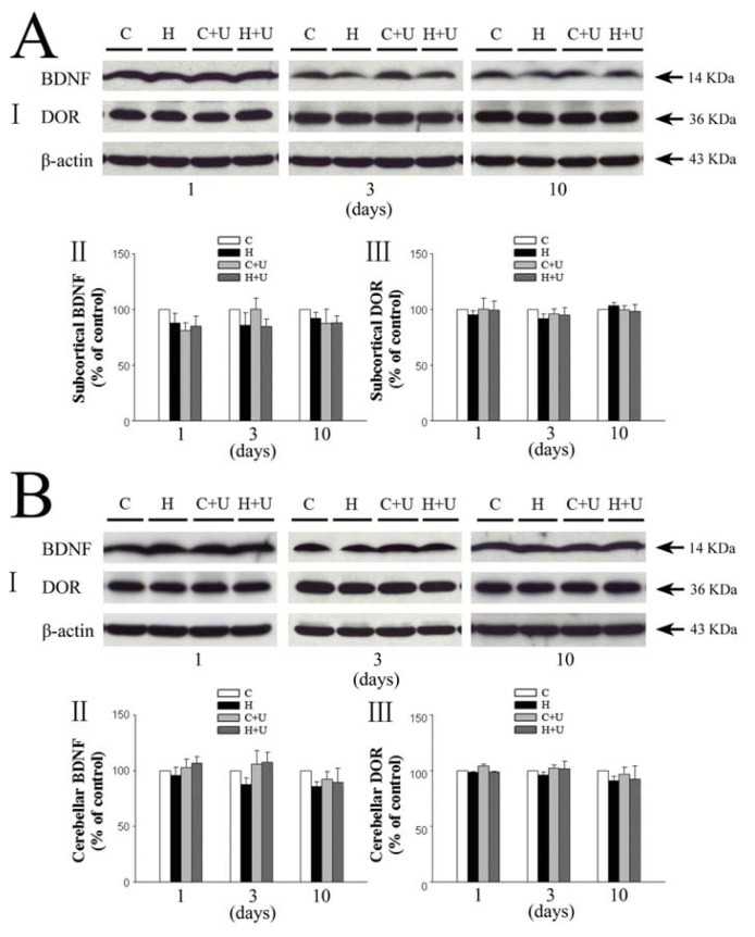
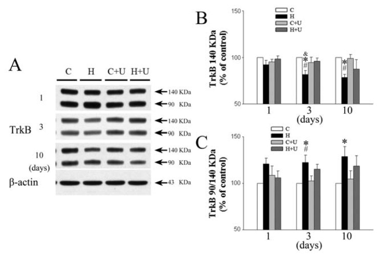
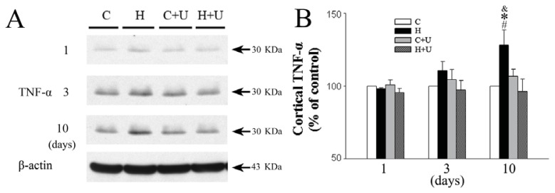
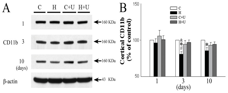
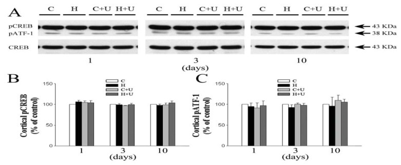
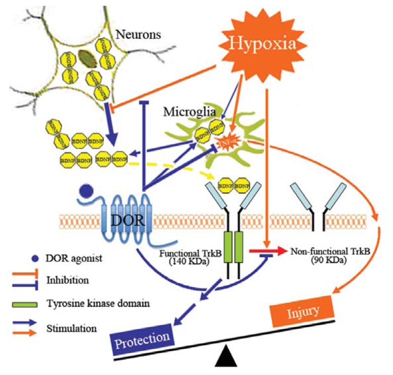
We investigated whether δ-opioid receptor (DOR)-induced neuroprotection involves the brain-derived neurotrophic factor (BDNF) pathway. We studied the effect of DOR activation on the expression of BDNF and other proteins in the cortex of C57BL/6 mice exposed to hypoxia (10% of oxygen) for 1-10 days. The results showed that: (1) 1-day hypoxia had no appreciable effect on BDNF expression, while 3- and 10-day hypoxia progressively decreased BDNF expression, resulting in 37.3% reduction (p < 0.05) after 10-day exposure; (2) DOR activation with UFP-512 (1 mg/kg, i.p., daily) partially reversed the hypoxia-induced reduction of BDNF expression in the 3- or 10-day exposed cortex; (3) DOR activation partially reversed the hypoxia-induced reduction in functional TrkB (140-kDa) and attenuated hypoxia-induced increase in truncated TrkB (90-kDa) in the 3- or 10-day hypoxic cortex; and (4) prolonged hypoxia (10 days) significantly increased TNF-α level and decreased CD11b expression in the cortex, which was completely reversed following DOR activation; and (5) there was no significant change in pCREB and pATF-1 levels in the hypoxic cortex. We conclude that prolonged hypoxia down-regulates BDNF-TrkB signaling leading to an increase in TNF-α in the cortex, while DOR activation up-regulates BDNF-TrkB signaling thereby decreasing TNF-α levels in the hypoxic cortex.


Figures

Figure 1
Figure 1
Effect of DOR activation on the expression of BDNF and DOR in the cortex and hippocampus exposed to hypoxia. (A) Effect of DOR activation on cortical expression of BDNF and DOR in hypoxia. I, Representative blots of Western blot analysis of the cortical tissues. II, Relative levels of BDNF. III, Relative levels of DOR. C, Normoxic control. H, Hypoxia. C + U (UFP-512), DOR activation with UFP-512 under normoxic conditions. H + U (UFP-512), DOR activation with UFP-512 under hypoxia. N = 4, 5 and 6 for 1, 3 and 10 days hypoxic exposure groups, respectively. * p < 0.05 vs. the control. #p < 0.05 vs. C + U (UFP-512). &p < 0.05 vs. H + U. Note that prolonged (3–10 days), but not relatively short-term (1 day), hypoxia significantly reduced the expression of BDNF but not of DOR, Furthermore, DOR activation with UFP-512 partially reversed the hypoxic reduction of BDNF expression; (B) Effect of DOR activation on hippocampal expression of BDNF and DOR in hypoxia. I, Representative blots of Western blot analysis of the cortical tissues. II, Relative levels of BDNF. III, Relative levels of DOR. C, Normoxic control. H, Hypoxia. C + U (UFP-512), DOR activation with UFP-512 in normoxic condition. H + U (UFP-512), DOR activation with UFP-512 in hypoxic condition. N = 4, 5 and 6 for 1, 3 and 10 days groups, respectively. * p < 0.05 vs. the control. Note that 1 day hypoxia significantly reduced the expression of BDNF in the hippocampus, while DOR activation partially reversed this hypoxic effect.
Figure 2
Figure 2
Effect of DOR activation on the expression of BDNF and DOR in the subcortical and cerebellar regions exposed to prolonged hypoxia. (A) Effect of DOR activation on BDNF and DOR expression in the subcortical region in hypoxia. I, Representative blots of Western blot analysis of the cortical tissues. II, Relative levels of BDNF. III, Relative levels of DOR. C, Normoxic control. H, Hypoxia. C + U (UFP-512), DOR activation with UFP-512 in normoxic condition. H + U (UFP-512), DOR activation with UFP-512 in hypoxic condition. Note that neither hypoxia nor DOR activation had any significant effect on the expression of BDNF and DOR in the subcortical region; (B) Effect of DOR activation on cerebellar expression of BDNF and DOR in hypoxia. I, Representative blots of Western blot analysis of the cortical tissues. II, Relative levels of BDNF. III, Relative levels of DOR. C, Normoxic control. H, Hypoxia. C + U (UFP-512), DOR activation with UFP-512 treatment under normoxic conditions. H + U (UFP-512), DOR activation with UFP-512 treatment under hypoxic conditions. N = 4, 5 and 6 for 1, 3 and 10 days groups, respectively. Note that neither hypoxia nor DOR activation had any significant effect on the expression of BDNF and DOR in the cerebellum.
Figure 3
Figure 3
Effect of DOR activation on cortical TrkB expression in hypoxia. (A) Representative blots of Western blot analysis of the cortical tissues; (B) Relative levels of 140 kDa TrkB. C, Relative levels of 90/140 kDa TrkB; (C) Normoxic control. H, Hypoxia. C + U (UFP-512), DOR activation with UFP-512 treatment under normoxic conditions. H + U (UFP-512), DOR activation with UFP-512 treatment under hypoxic conditions. N = 4, 5 and 6 for 1, 3 and 10 days groups, respectively. * p < 0.05 vs. the control. #p < 0.05 vs. C + U (UFP-512). &p < 0.05 vs. H + U (UFP-512). Note that 3 or 10 days hypoxia significantly reduced the level of 140 KDa TrkB and increased that of 90/140 kDa TrkB in the cortex, while DOR activation largely reversed these hypoxia-induced changes.
Figure 4
Figure 4
Effect of DOR activation on cortical TNF-α expression in hypoxia. (A) Representative blots of Western blot analysis of the cortical tissues; (B) Relative levels of TNF-α. C, Normoxic control. H, Hypoxia. C + U (UFP-512), DOR activation with UFP-512 in normoxic condition. H + U (UFP-512), DOR activation with UFP-512 in hypoxic condition. N = 4, 5 and 6 for 1, 3 and 10 days groups, respectively. * p < 0.05 vs. the control. #p < 0.05 vs. C + U (UFP-512). &p < 0.05 vs. H + U (UFP-512). Note that 10 days hypoxia significantly increased the level of TNF-α, while DOR activation completely inhibited such increase.
Figure 5
Figure 5
Effect of DOR activation on cortical CD11b expression in hypoxia. (A) Representative blots of Western blot analysis of the cortical tissues; (B) Relative levels of CD11b. C, Normoxic control. H, Hypoxia. C + U (UFP-512), DOR activation with UFP-512 treatment under normoxic conditions. H + U (UFP-512), DOR activation with UFP-512 treatment under hypoxic conditions. N = 4, 5 and 6 for 1, 3 and 10 days groups, respectively. * p < 0.05 vs. the control. #p < 0.05 vs. C + U (UFP-512). &p < 0.05 vs. H + U (UFP-512). Note that 3 or 10 days hypoxia significantly reduced the level of CD11b, while DOR activation with UFP-512 reversed this hypoxic reduction.
Figure 6
Figure 6
Effect of DOR activation on cortical pCREB and pATF-1 expression in hypoxia. (A) Representative blots of Western blot analysis of the cortical tissues; (B) Relative levels of pCREB. C, Relative levels of pATF-1; (C) Normoxic control. H, Hypoxia. C + U (UFP-512), DOR activation with UFP-512 treatment under normoxic conditions. H + U (UFP-512), DOR activation with UFP-512 treatment under hypoxic conditions. N = 4, 5 and 6 for 1, 3 and 10 days groups, respectively. Note that neither hypoxia nor DOR activation had any significant effect on the expression of pCREB and pATF-1 in the cortex.
Figure 7
Figure 7
Schematic demonstration of the DOR-BDNF-TrkB pathway in brain protection against hypoxic injury. Prolonged hypoxia causes a down-regulation of BDNF-TrkB signals resulting in an increased cortical TNF-α, while DOR activation up-regulates BDNF-TrkB signals thereby decreasing the level of TNF-α in the hypoxic cortex.

References

    1. Zhang J., Haddad G.G., Xia Y. Delta-, but not mu- and kappa-, opioid receptor activation protects neocortical neurons from glutamate-induced excitotoxic injury. Brain Res. 2000;885:143–153. - PubMed
    1. Tian X.S., Zhou F., Yang R., Xia Y., Wu G.C., Guo J.C. Effects of intracerebroventricular injection of delta-opioid receptor agonist TAN-67 or antagonist naltrindole on acute cerebral ischemia in rat. Sheng Li Xue Bao. 2008;60:475–484. - PubMed
    1. He X., Sandhu H.K., Yang Y., Hua F., Belser N., Kim D.H., Xia Y. Neuroprotection against hypoxia/ischemia: Delta-opioid receptor-mediated cellular/molecular events. Cell Mol. Life Sci. 2013;70:2291–2303. - PMC - PubMed
    1. Zhao P., Huang Y., Zuo Z. Opioid preconditioning induces opioid receptor-dependent delayed neuroprotection against ischemia in rats. J. Neuropathol. Exp. Neurol. 2006;65:945–952. - PubMed
    1. Gwak M.S., Li L., Zuo Z. Morphine preconditioning reduces lipopolysaccharide and interferon-gamma-induced mouse microglial cell injury via delta 1 opioid receptor activation. Neuroscience. 2010;167:256–260. - PMC - PubMed

Publication types

MeSH terms

Substances

LinkOut - more resources