. 2013 Oct;16(4):985-92.
doi: 10.1007/s10456-013-9374-5. Epub 2013 Aug 4.

Neuronal sirtuin1 mediates retinal vascular regeneration in oxygen-induced ischemic retinopathy

Affiliations

Neuronal sirtuin1 mediates retinal vascular regeneration in oxygen-induced ischemic retinopathy

Jing Chen et al. Angiogenesis. 2013 Oct.

Abstract

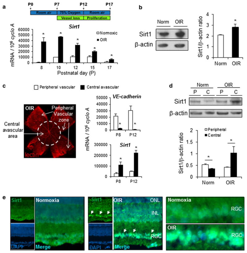
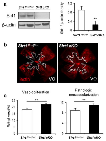
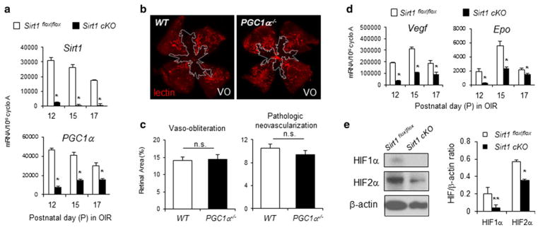
Regeneration of blood vessels in ischemic neuronal tissue is critical to reduce tissue damage in diseases. In proliferative retinopathy, initial vessel loss leads to retinal ischemia, which can induce either regrowth of vessels to restore normal metabolism and minimize damage, or progress to hypoxia-induced sight-threatening pathologic vaso-proliferation. It is not well understood how retinal neurons mediate regeneration of vascular growth in response to ischemic insults. In this study we aim to investigate the potential role of Sirtuin 1 (Sirt1), a metabolically-regulated protein deacetylase, in mediating the response of ischemic neurons to regulate vascular regrowth in a mouse model of oxygen-induced ischemic retinopathy (OIR). We found that Sirt1 is highly induced in the avascular ischemic retina in OIR. Conditional depletion of neuronal Sirt1 leads to significantly decreased retinal vascular regeneration into the avascular zone and increased hypoxia-induced pathologic vascular growth. This effect is likely independent of PGC-1α, a known Sirt1 target, as absence of PGC-1α in knockout mice does not impact vascular growth in retinopathy. We found that neuronal Sirt1 controls vascular regrowth in part through modulating deacetylation and stability of hypoxia-induced factor 1α and 2α, and thereby modulating expression of angiogenic factors. These results indicate that ischemic neurons induce Sirt1 to promote revascularization into ischemic neuronal areas, suggesting a novel role of neuronal Sirt1 in mediating vascular regeneration in ischemic conditions, with potential implications beyond retinopathy.


Conflict of interest statement

Conflict of interest DAS is a consultant to and inventor on patents licensed to GlaxoSmithKline and Ovascience.

Figures

Fig. 1
Fig. 1
Sirt1 is induced in avascular retina in a mouse model of oxygen-induced retinopathy (OIR). a Sirt1 mRNA expression is significantly upregulated in a mouse model of oxygen-induced retinopathy, in which neonatal pups are exposed to 75 % oxygen from postnatal day (P) 7 to P12. b Sirt1 protein levels are upregulated in OIR retinas compared to normoxic controls (Norm) at P8. c At P12 in OIR, retinal vasculature is visualized with lectin staining (red) showing central avascular zone and peripheral vascular zones (arrows), which were separated by micro-dissection. Sirt1 and VE-cadherin mRNA expression levels were quantified with RT-qPCR in peripheral vascular versus central avascular samples isolated at P8 and P12 in OIR. Central avascular retina samples show a lack of vascular endothelial cell marker VE-cadherin, confirming absence of vessels. Sirt1 is significantly up-regulated in central avascular retinal samples compared to samples from peripheral vascular region. d Western blot with Sirt1 antibody shows upregulated Sirt1 protein levels in samples isolated from central avascular regions of retinas in OIR compared with peripheral vascular regions at P12. In room air control retinas (Norm) not exposed to OIR, Sirt1 levels are lowered in the central region (C) compared to peripheral retinal region (P). e Immunohistochemistry staining of retinal cross sections from P17 OIR mouse or room air control shows increased staining of Sirt1 antibody (green) in the retinal ganglion cell layer (arrows), with enrichment in the cellular nucleus region visualized by double staining of DAPI (blue). ONL outer nuclear layer, INL inner nuclear layer, RGC retinal ganglion cell layer. *p ≤ 0.05. (Color figure online)
Fig. 2
Fig. 2
Depletion of neuronal Sirt1 decreases retinal revascularization in oxygen-induced retinopathy and consequently precipitates pathologic neovascularization. a Western blots show that Sirt1 protein levels are significantly decreased in the retina of neuronal specific conditional Sirt1 knockout mice (Sirt1 cKO) compared to littermate control mice (Sirt1flox|flox). b Representative images of retinal vasculature from P17 Sirt1 cKO mice and littermate control Sirt1flox|flox mice with induced retinopathy (OIR). Blood vessels were stained with lectin (red) with areas of vaso-obliteration highlighted by white line. Quantification of retinopathy in Sirt1 cKO and control retina with OIR shows significantly increased levels of vaso-obliteration (absence of red) and pathologic neovascularization (bright red) (n = 18–20 per group). **p ≤ 0.01. (Color figure online)
Fig. 3
Fig. 3
Sirt1 regulation of vascular regrowth in retinopathy is likely mediated via regulation of hypoxia growth factors (HIF) 1α and 2α, other than PGC-1α. a At P12, 15, and 17 in OIR, depletion of neuronal Sirt1 results in significantly decreased mRNA levels of Sirt1 and PGC-1a in the retinas compared to littermate Sirt1flox|flox controls. *p ≤ 0.05. b PGC-1a−/− mice do not show significant differences in vaso-obliteration (outlined in white) and pathologic neovascularization in OIR at P17 compared to littermate WT controls (n = 10–11 per group, n.s. not significant). d OIR exposed Sirt1 cKO retinas show significantly decreased mRNA levels of angiogenic factors Vegf and Epo in the retinas compared to littermate Sirt1flox|flox controls. *p ≤ 0.05. e Western blot with Sirt1 antibody shows decreased levels of HIF1α and 2α protein in Sirt1 cKO retinas compared to littermate Sirt1flox|flox control retinas
Fig. 4
Fig. 4
Sirt1 regulates acetylation of HIF1α and 2α, and expression of Epo in RGC-5 culture. a RGC-5 cells differentiated after incubation with staurosporine show dendrites and axons (arrows) characteristic of neurons. RGC-5 cells also show staining with antibody against βIII-tubulin (green), a RGC marker. b Incubation of RGC-5 cells with Ex527, a specific Sirt1 inhibitor, results in increased levels of protein acetylation detected by an acetyl-lysine antibody. c Inhibition of Sirt1 with Ex527 in RGC-5 culture increased protein acetylation of HIF1α and HIF2α, detected by immunoprecipitation with acetyl-lysine antibody and followed by Western blots against HIF1α or HIF2α. d Ex527 treatment of RGC-5 cells leads to decreased expression level of Epo mRNA measured by RT-qPCR, and decreased Epo protein level measured by ELISA assay. e Aortic ring explants cultured with conditioned medium from RGC-5 culture treated with Sirt1 inhibitor Ex527 shows significantly decreased levels of vascular growth compared with RGC-5 medium treated with vehicle control (vascular sprouts outlined in white, n = 6–10 per group). f Depletion of Sirt1 in neuronal retina leads to increased mRNA expression of inflammatory cytokines TNFα, IL-1β, IL-6 and decreased mRNA level of SOCS3 at P12 in OIR. g Depletion of Sirt1 in neuronal retinal results in increased mRNA expression of anti-angiogenic factors TSP-1, TSP-2, TIMP-1 and PEDF at P12 in OIR. *p ≤ 0.05; **p ≤ 0.01. (Color figure online)

References

    1. Antonetti DA, et al. Diabetic retinopathy. N Engl J Med. 2012;366:1227–1239. - PubMed
    1. Sapieha P. Eyeing central neurons in vascular growth and reparative angiogenesis. Blood. 2012;120:2182–2194. - PubMed
    1. Akula JD, et al. The neurovascular relation in oxygen-induced retinopathy. Mol Vis. 2008;14:2499–2508. - PMC - PubMed
    1. Sapieha P, et al. The succinate receptor GPR91 in neurons has a major role in retinal angiogenesis. Nat Med. 2008;14:1067–1076. - PubMed
    1. Fukushima Y, et al. Sema3E-PlexinD1 signaling selectively suppresses disoriented angiogenesis in ischemic retinopathy in mice. J Clin Invest. 2011;121:1974–1985. - PMC - PubMed

Publication types

MeSH terms

Substances

LinkOut - more resources