. 2013 Jul 31;18(8):9195-206.
doi: 10.3390/molecules18089195.

Inhibition of lipopolysaccharide-induced proinflammatory responses by Buddleja officinalis extract in BV-2 microglial cells via negative regulation of NF-kB and ERK1/2 signaling

Affiliations

Inhibition of lipopolysaccharide-induced proinflammatory responses by Buddleja officinalis extract in BV-2 microglial cells via negative regulation of NF-kB and ERK1/2 signaling

Won-Jun Oh et al. Molecules. .

Abstract

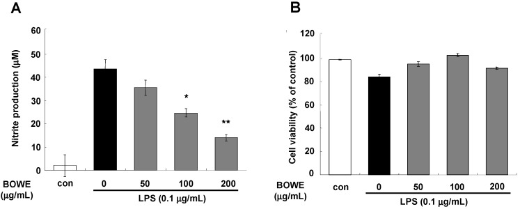
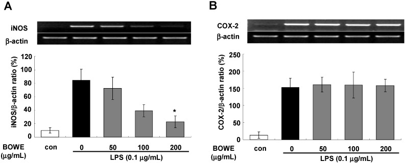
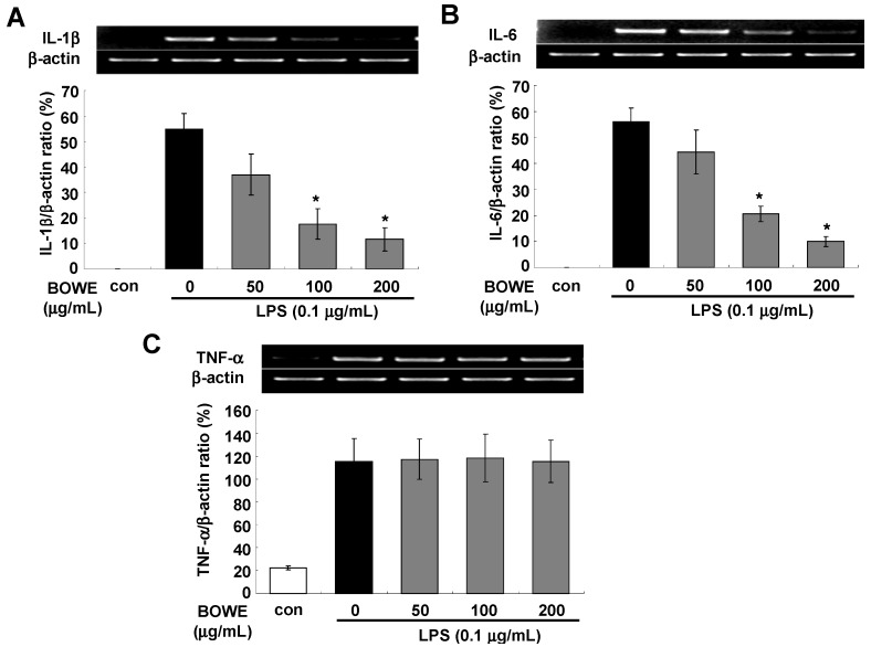
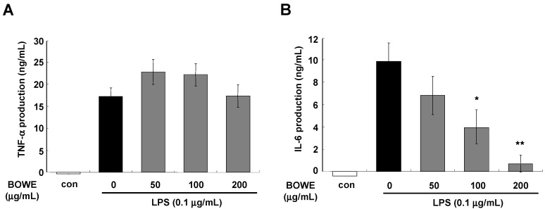
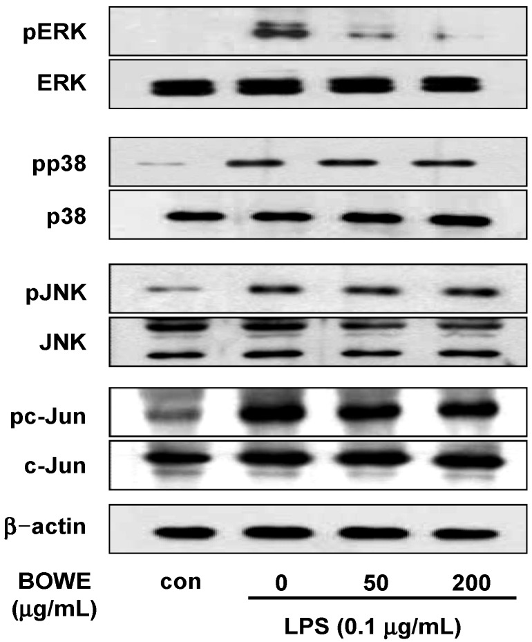
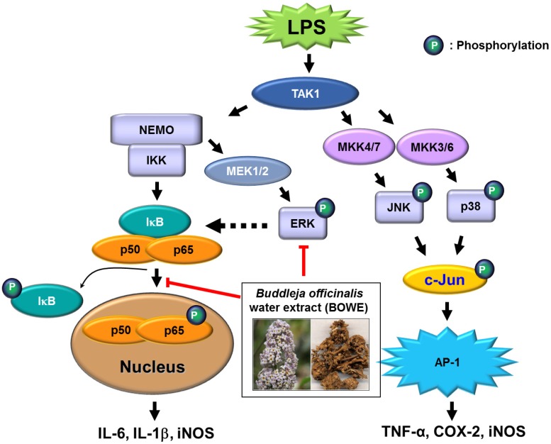
Buddleja officinalis has been traditionally used in the supportive treatment of inflammatory and neuronal diseases in Korea and China. Although several reports have shown the anti-inflammatory effects of Buddleja officinalis, the anti-neuroinflammatory effect has remained unclear. In this study, we aimed to investigate the inhibitory effects of flower buds of B. officinalis Maximowicz water extract (BOWE) on LPS-induced inflammatory processes in BV-2 microglial cells. BOWE dose-dependently inhibited the production of nitric oxide as well as iNOS mRNA expression. Moreover, BOWE prevented IL-1β and IL-6 mRNA expression. However, BOWE had no effect on LPS-induced COX-2 or TNF-a mRNA expression. The extract also had no effect on LPS-stimulated p38 MAPK, JNK, and c-Jun phosphorylation, whereas ERK1/2 phosphorylation was strongly inhibited by BOWE. BOWE also inhibited the LPS-induced degradation of IkB-α, and LPS-induced phosphorylation of p65 NF-kB protein. These data indicate that BOWE inhibited the nitric oxide production and pro-inflammatory gene expression in BV-2 microglial cells, possibly through a negative regulation of the NF-kB and ERK1/2 pathways. Further identification of the direct target molecule(s) of BOWE is required to support its use as an anti-neuroinflammatory agent against the neurodegenerative disorders.


Figures

Figure 1
Figure 1
Effect of BOWE on LPS-induced NO (A) and cell viability assay (B): BV-2 cells were pretreated with or without the indicated doses of BOWE for 4 h and then stimulated with LPS (0.1 μg/mL) for 18 h. The culture supernatants were subsequently collected and analyzed for nitrite levels. The cell viability was determined by CCK assay. Data are presentedas the means ± SEM of triplicates from separate experiments. * p < 0.05 and ** p < 0.01 denote significant differences compared to the group treated with LPS only.
Figure 2
Figure 2
Effect of BOWE on LPS-induced mRNA expression levels of iNOS and COX-2 in BV-2 cells: For mRNA expression, BV-2 cells were pretreated with BOWE for 4 h and then stimulated with LPS (0.1 μg/mL) for 18 h. Total RNA was isolated, and mRNA levels of iNOS (A) and COX-2 (B) were then measured by RT-PCR. β actin expression was used as an internal control. The quantification of relative band intensities from three independent experiments were analyzed by densitometry and expressed as means ± SEM. * p < 0.01 denotes a significant difference compared to the group treated with LPS only.
Figure 3
Figure 3
Effect of BOWE on the mRNA expressions of IL-1β (A), IL-6 (B), and TNF-α (C): BV-2 cells were pretreated with or without the indicated concentrations of BOWE for 4 h and then stimulated with LPS (0.1 μg/mL) for 18 h. Total RNA was isolated and mRNA expressions of the aforementioned cytokines were measured by RT-PCR. β actin was used as a housekeeping gene. The quantification of relative band intensities from three independentexperiments were analyzed by densitometry and expressed as means ± SEM. * p < 0.01denotes a significant difference compared to the group treated with LPS only.
Figure 4
Figure 4
Effect of BOWE on LPS-induced TNF-α (A) and IL-6 (B) release: BV-2 cells were pretreated with or without the indicated concentrations of BOWE for 4 h and then stimulated with LPS (0.1 μg/mL) for 18 h. The culture supernatants were subsequently harvested and analyzed for TNF-α and IL-6 levels by ELISA. Data are presented as the means ± SEM in each group (n = 3). * p < 0.05 and ** p < 0.01 denote significant differences compared to the group treated with LPS only.
Figure 5
Figure 5
Effect of BOWE on IκB-α and NF-κB p65 phosphorylation in LPS-stimulated BV-2 cells: BV-2 cells were pretreated with or without the indicated concentrations of BOWE for 4 h and then stimulated with LPS (0.1 μg/mL) for 18 h. The cellular proteins from the cells were used for the detection of phosphorylated forms of IκB-α and NF-κB p65 by western blotting. β-actin was used as a loading control.
Figure 6
Figure 6
Effect of BOWE on MAPK phosphorylation in LPS-stimulated BV-2 cells: BV-2 cells were pretreated with or without the indicated concentrations of BOWE for 4 h and then stimulated with LPS (0.1 μg/mL) for 18 h. The cellular proteins from the cells were used for the detection of phosphorylated or total forms of ERK1/2, p38 MAPK, JNK1/2, and c-Jun by western blotting.
Figure 7
Figure 7
Suggested anti-inflammation mechanism of BOWE in LPS-treated BV-2 cells: In BV-2 cells treated with LPS, BOWE prevented the activation of ERK1/2 and NF-κB, and subsequently attenuated the production of IL-6, IL-1β, iNOS, and nitric oxide. BOWE did not have any effect on the activation of JNK, p38, and c-Jun and the production of TNF-α and COX-2. The negative regulation of ERK1/2 and NF-κB activation is supposed to be the main mechanism of anti-inflammatory action of BOWE in BV-2 cells.

References

    1. Whitney N.P., Eidem T.M., Peng H., Huang Y., Zheng J.C. Inflammation mediates varying effects in neurogenesis: relevance to the pathogenesis of brain injury and neurodegenerative disorders. J. Neurochem. 2009;108:1343–1359. doi: 10.1111/j.1471-4159.2009.05886.x. - DOI - PMC - PubMed
    1. Liu B. Role of Microglia in Inflammation-Mediated Neurodegenerative Diseases: Mechanisms and Strategies for Therapeutic Intervention. J. Pharm. Exp. Ther. 2003;304:1–7. doi: 10.1124/jpet.102.035048. - DOI - PubMed
    1. Saha R.N., Pahan K. Regulation of Inducible Nitric Oxide Synthase Gene in Glial Cells. Antioxid. Redox Signal. 2006;8:929–947. doi: 10.1089/ars.2006.8.929. - DOI - PMC - PubMed
    1. Kim Y.S., Joh T.H. Microglia, major player in the brain inflammation: Their roles in the pathogenesis of Parkinson’s disease. Exp. Mol. Med. 2006;38:333–347. doi: 10.1038/emm.2006.40. - DOI - PubMed
    1. McGeer E.G., McGeer P.L. Inflammatory processes in Alzheimer’s disease. Prog. Neuro Psychoph. 2003;27:741–749. doi: 10.1016/S0278-5846(03)00124-6. - DOI - PubMed

Publication types

MeSH terms

LinkOut - more resources