. 2013 Dec 20;22(25):5107-20.
doi: 10.1093/hmg/ddt365. Epub 2013 Aug 2.

Epigenetic regulation of COL15A1 in smooth muscle cell replicative aging and atherosclerosis

Affiliations

Epigenetic regulation of COL15A1 in smooth muscle cell replicative aging and atherosclerosis

Jessica J Connelly et al. Hum Mol Genet. .

Abstract

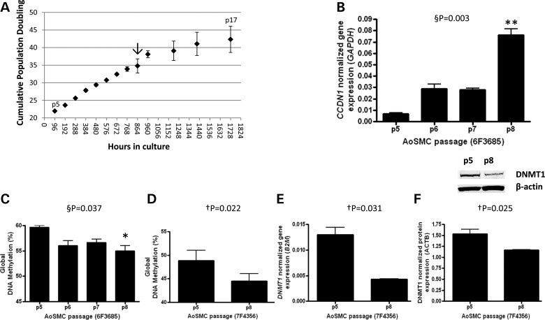
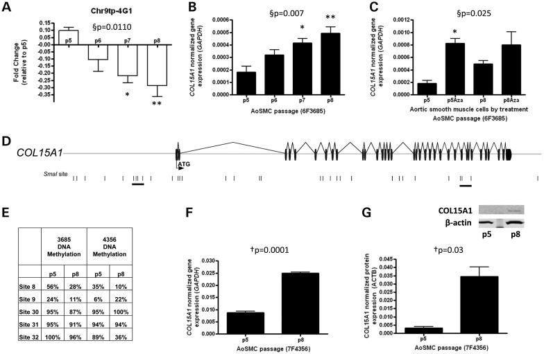
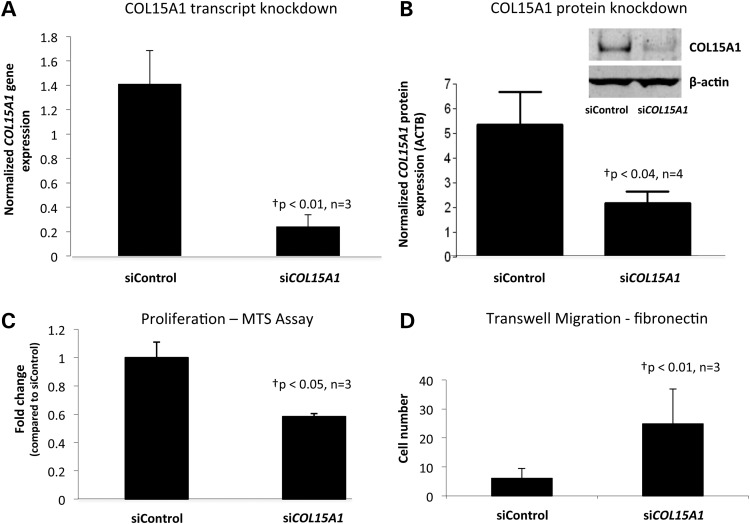
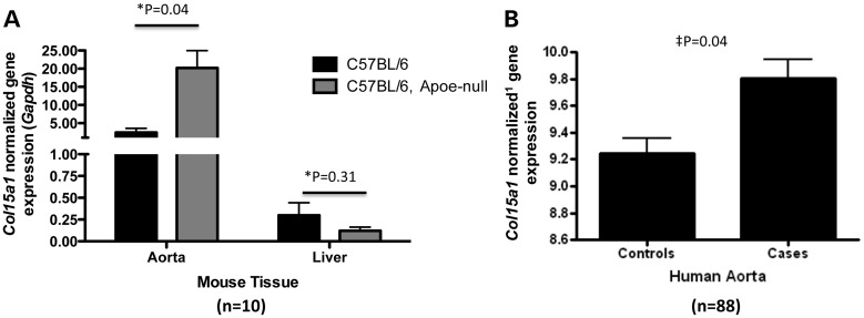
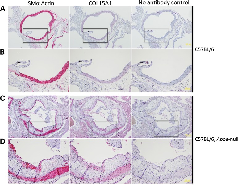
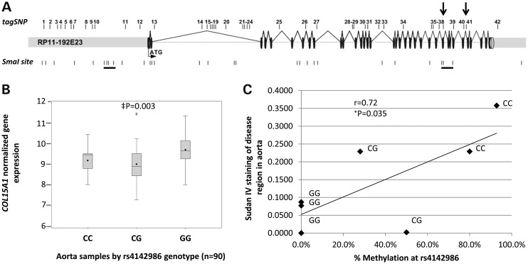
Smooth muscle cell (SMC) proliferation is a hallmark of vascular injury and disease. Global hypomethylation occurs during SMC proliferation in culture and in vivo during neointimal formation. Regardless of the programmed or stochastic nature of hypomethylation, identifying these changes is important in understanding vascular disease, as maintenance of a cells' epigenetic profile is essential for maintaining cellular phenotype. Global hypomethylation of proliferating aortic SMCs and concomitant decrease of DNMT1 expression were identified in culture during passage. An epigenome screen identified regions of the genome that were hypomethylated during proliferation and a region containing Collagen, type XV, alpha 1 (COL15A1) was selected by 'genomic convergence' for characterization. COL15A1 transcript and protein levels increased with passage-dependent decreases in DNA methylation and the transcript was sensitive to treatment with 5-Aza-2'-deoxycytidine, suggesting DNA methylation-mediated gene expression. Phenotypically, knockdown of COL15A1 increased SMC migration and decreased proliferation and Col15a1 expression was induced in an atherosclerotic lesion and localized to the atherosclerotic cap. A sequence variant in COL15A1 that is significantly associated with atherosclerosis (rs4142986, P = 0.017, OR = 1.434) was methylated and methylation of the risk allele correlated with decreased gene expression and increased atherosclerosis in human aorta. In summary, hypomethylation of COL15A1 occurs during SMC proliferation and the consequent increased gene expression may impact SMC phenotype and atherosclerosis formation. Hypomethylated genes, such as COL15A1, provide evidence for concomitant epigenetic regulation and genetic susceptibility, and define a class of causal targets that sit at the intersection of genetic and epigenetic predisposition in the etiology of complex disease.


Figures

Figure 1.
Figure 1.
Proliferating aortic SMCs exhibit early molecular signs of senescence. (A) Aortic SMCs were grown in culture in triplicate under proliferating conditions and passaged when cells reached 80% confluence. PD with time was calculated. Arrow indicates first change in cell growth between 35 and 40 PDs. (B) Cyclin D1 (CCDN1) transcript levels were assayed by passage to assess changes in the senescent state. (C) Global DNA methylation level was measured by a LUMA assay by passage and (D) in a second AoSMC line for validation. (E) DNMT1 transcript level and (F) protein level was measured in the AoSMC validation line. A representative western blot is shown for DNMT1 and loading control β-actin. §P, repeated measures ANOVA, Bonferroni's multiple comparison test *P < 0.05, **P < 0.01; P, paired t-test, two-tailed.
Figure 2.
Figure 2.
Identification of COL15A1 as a gene that is epigenetically regulated in smooth muscle replicative aging. (A) The clone containing COL15A1 exhibits decreased methylation with passage. This was assessed by microarray hybridization of p5, p6, p7 and p8 methylated DNA and displayed as fold change (log2[p8/p5]). (B) COL15A1 gene expression was measured by RT-PCR with age of SMC (n = 3) and normalized to glyceraldehyde-3-phosphate dehydrogenase (GAPDH). (C) Normalized COL15A1 gene expression was measured by RT-PCR in both young and aged cells in the absence and presence of a demethylating chemical, 5-Aza-2′-deoxycytidine (Aza). Data were normalized to GAPDH. (D) A schematic of the clone containing COL15A1 identified in a screen for genes that change DNA methylation with SMC age. The start codon (ATG) is contained within the first exon; exons are denoted as black discs and introns as lines. SmaI sites contained within this region are denoted by a black vertical line. Groups of SmaI sites that are hypomethylated with age are underlined. (E) Bisulfite cloned sequencing results from two passaged AoSMCs lines. Indicated sites are part of the two underlined clusters in the gene schematic. (F) Validation of COL15A1 expression changes with passage in second AoSMC line (7F4356). (G) Total cellular COL15A1 protein levels were assayed in young (p5) and aged (p8) SMCs by western blot. Quantitation (n = 3) was performed on a LI-COR Odyssey Infrared Imaging System. Data were normalized to β-actin (ACTB). A representative western blot is shown for COL15A1 and loading control β-actin. §P, repeated measures ANOVA, *Bonferroni's Multiple Comparison test P < 0.05; **P < 0.01. P, paired t-test (n = 3), two-tailed.
Figure 3.
Figure 3.
COL15A1 levels modify SMC phenotype. (A) Human AoSMCs (7F4356) were transfected with siCOL15A1 or siControl and the level of COL15A1 (A) gene expression normalized to Beta-2 Microglobulin (B2M) and (B) protein expression normalized to β-actin (ACTB) were measured. A representative western blot is shown for COL15A1 and loading control β-actin. (C) COL15A1 control or knockdown cells were assayed for proliferation using the MTS assay from Promega. Data were normalized to the siControl. (D) COL15A1 control and knockdown cells were assayed for their ability to migrate through a permeable membrane in response to fibronectin. P, paired t-test (n = 3), two-tailed.
Figure 4.
Figure 4.
COL15A1 expression increases with atherosclerosis. (A) Col15a1 mRNA levels were measured by RT-PCR in thoracic aorta or liver taken from wild type (C57/BL6) chow-fed mice (n = 5) or Apoe null mice (n = 5) after 13 weeks of a high fat Western diet feeding. Expression of Col15a1 in the thoracic aorta but not liver is significantly increased with accelerated atherosclerosis (high fat diet feeding in an Apoe null background). (B) Microarray gene expression data for COL15A1 from aorta tissue with disease (Cases, n = 26) or without disease (Controls, n = 62). Disease was quantitated using Sudan IV staining for lipids and by counting raised lesions. Controls showed neither lipid staining nor raised lesions. *P, unpaired t-test, two-tailed. P, mixed model (see methods). 1microarray data normalization provided in methods.
Figure 5.
Figure 5.
Col15a1 is expressed in SMCs in the mouse aorta. Transverse sections of the aortic root were taken from a wild-type C57BL/6 chow-fed mouse (A and B) or a mouse model of atherosclerosis (C57BL/6 Apoe null) fed a high fat Western diet (C and D). Eighteen weeks of Western diet feeding in the Apoe null background induced atherosclerosis (A versus C). Regions boxed in A and C are shown enlarged in B and D, respectively. Scale is indicated in yellow. Four micron sections were stained with an antibody to smooth muscle alpha actin (Acta2) or Col15a1.
Figure 6.
Figure 6.
A genetic variant in COL15A1 is associated with atherosclerosis, leads to reduced COL15A1 gene expression in human aorta and its methylation state is correlated with disease. (A) Forty-two tag SNPs were selected to cover all of the known genetic variation in COL15A1. These SNPs were genotyped in an atherosclerosis sample (CATHGEN) and allelic association with atherosclerosis was assessed using multivariable logistic regression modeling adjusting for sex and known CAD risk factors. Significant (SNP38) and trending (SNP 41) SNPs are indicated with arrows. Underlined SmaI sites indicate sites that change DNA methylation with passage. (B) COL15A1 gene expression was assessed in aorta tissue regardless of disease and the data were grouped by genotype. C allele carriers have a significant reduction in COL15A1 gene expression. (C) The C allele of rs4142986 is the minor allele and creates a putative DNA methylation site in a region of the gene that changes methylation with age. DNA methylation at rs4142986 was measured in aorta samples scored for disease (Sudan IV). DNA Methylation (%) is plotted versus disease for individuals of each genotype. There is a significant correlation with disease and increased methylation. P, mixed model (see methods); †P, Pearson's correlation, two-tailed.

References

    1. Owens G.K. Regulation of differentiation of vascular smooth muscle cells. Physiol. Rev. 1995;75:487–517. - PubMed
    1. Watkins H., Farrall M. Genetic susceptibility to coronary artery disease: from promise to progress. Nat. Rev. Genet. 2006;7:163–173. - PubMed
    1. Owens G.K., Kumar M.S., Wamhoff B.R. Molecular regulation of vascular smooth muscle cell differentiation in development and disease. Physiol. Rev. 2004;84:767–801. - PubMed
    1. Libby P., Geng Y.J., Sukhova G.K., Simon D.I., Lee R.T. Molecular determinants of atherosclerotic plaque vulnerability. Ann. N. Y. Acad. Sci. 1997;811:134–142. discussion 142–5. - PubMed
    1. Davies M.J., Woolf N., Rowles P., Richardson P.D. Lipid and cellular constituents of unstable human aortic plaques. Basic Res. Cardiol. 1994;89(Suppl 1):33–39. - PubMed

Publication types