

Quantification of cardiomyocyte hypertrophy by cardiac magnetic resonance: implications for early cardiac remodeling
- PMID: 23912910
- PMCID: PMC5308548
- DOI: 10.1161/CIRCULATIONAHA.112.000438
Quantification of cardiomyocyte hypertrophy by cardiac magnetic resonance: implications for early cardiac remodeling
Abstract
Background: Cardiomyocyte hypertrophy is a critical precursor to the development of heart failure. Methods to phenotype cellular hypertrophy noninvasively are limited. The goal was to validate a cardiac magnetic resonance-based approach for the combined assessment of extracellular matrix expansion and cardiomyocyte hypertrophy.
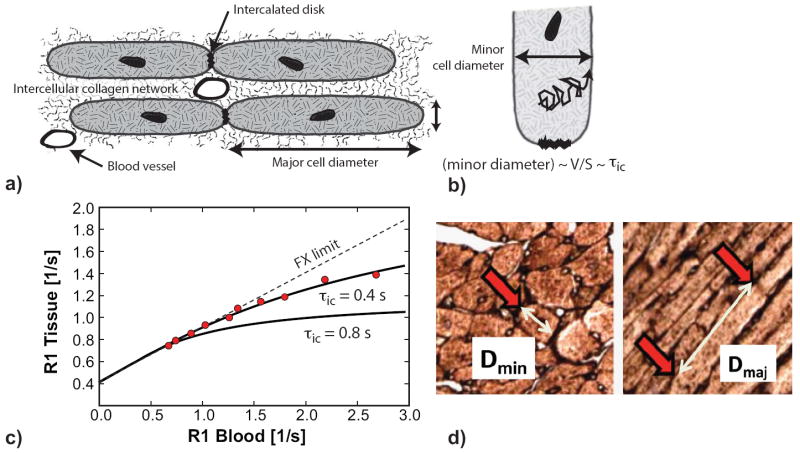
Methods and results: Two murine models of hypertension (n=18, with n=15 controls) induced by l-N(G)-nitroarginine methyl ester (L-NAME) and pressure overload (n=11) from transaortic constriction (TAC) were imaged by cardiac magnetic resonance at baseline and 7 weeks after L-NAME treatment or up to 7 weeks after TAC. T1 relaxation times were measured before and after gadolinium contrast. The intracellular lifetime of water (τic), a cell size-dependent parameter, and extracellular volume fraction, a marker of interstitial fibrosis, were determined with a model for transcytolemmal water exchange. Cardiomyocyte diameter and length were measured on FITC-wheat germ agglutinin-stained sections. The τic correlated strongly with histological cardiomyocyte volume-to-surface ratio (r=0.78, P<0.001) and cell volume (r=0.75, P<0.001). Histological cardiomyocyte diameters and cell volumes were higher in mice treated with L-NAME compared with controls (P<0.001). In the TAC model, cardiac magnetic resonance and histology showed cell hypertrophy at 2 weeks after TAC without significant fibrosis at this early time point. Mice exposed to TAC demonstrated a significant, longitudinal, and parallel increase in histological cell volume, volume-to-surface ratio, and τic between 2 and 7 weeks after TAC.
Conclusion: The τic measured by contrast-enhanced cardiac magnetic resonance provides a noninvasive measure of cardiomyocyte hypertrophy. Extracellular volume fraction and τic can track myocardial tissue remodeling from pressure overload.
Keywords: hypertrophy; magnetic resonance imaging.
Conflict of interest statement
Disclosures: None.
Figures


References
-
- Drazner MH. The progression of hypertensive heart disease. Circulation. 2011;123:327–334. - PubMed
-
- Devereux RB, Dahlof B, Levy D, Pfeffer MA. Comparison of enalapril versus nifedipine to decrease left ventricular hypertrophy in systemic hypertension (the preserve trial) Am J Cardiol. 1996;78:61–65. - PubMed
-
- Dweck MR, Joshi S, Murigu T, Gulati A, Alpendurada F, Jabbour A, Maceira A, Roussin I, Northridge DB, Kilner PJ, Cook SA, Boon NA, Pepper JR, Mohiaddin RH, Newby DE, Pennell DJ, Prasad SK. Left ventricular remodelling and hypertrophy in patients with aortic stenosis: Insights from cardiovascular magnetic resonance. J Cardiovasc Magn Reson. 2012;14:50. - PMC - PubMed
-
- Sharma S, Maron BJ, Whyte G, Firoozi S, Elliott PM, McKenna WJ. Physiologic limits of left ventricular hypertrophy in elite junior athletes: Relevance to differential diagnosis of athlete’s heart and hypertrophic cardiomyopathy. J Am Coll Cardiol. 2002;40:1431–1436. - PubMed
Publication types
MeSH terms
Substances
Grants and funding
LinkOut - more resources
Full Text Sources
Other Literature Sources
Medical
