. 2013 Sep;16(9):1238-47.
doi: 10.1038/nn.3479. Epub 2013 Aug 4.

Top3β is an RNA topoisomerase that works with fragile X syndrome protein to promote synapse formation

Affiliations

Top3β is an RNA topoisomerase that works with fragile X syndrome protein to promote synapse formation

Dongyi Xu et al. Nat Neurosci. 2013 Sep.

Abstract

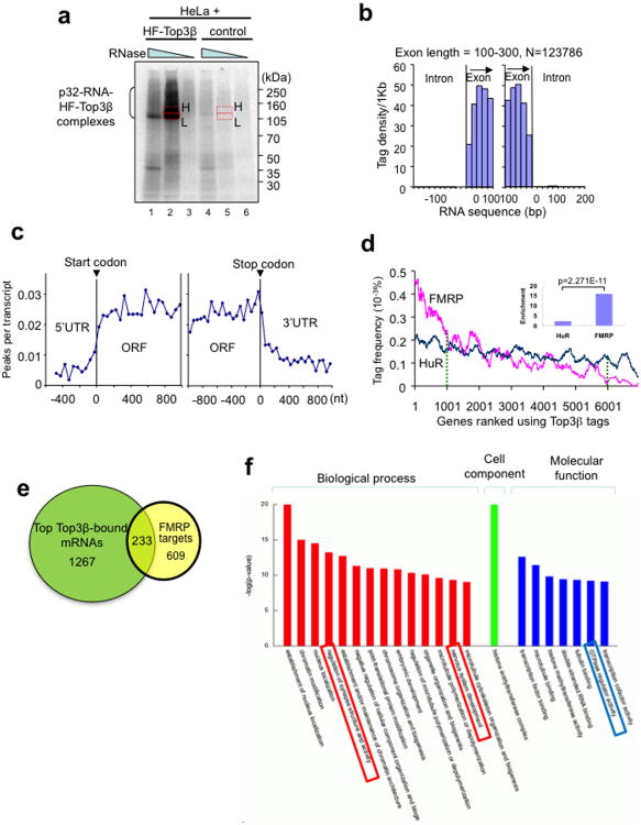
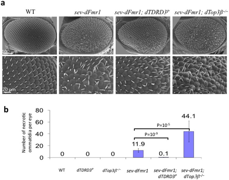
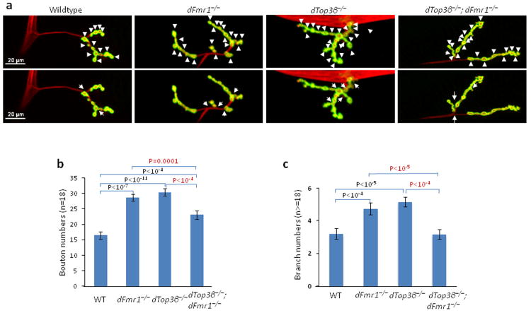
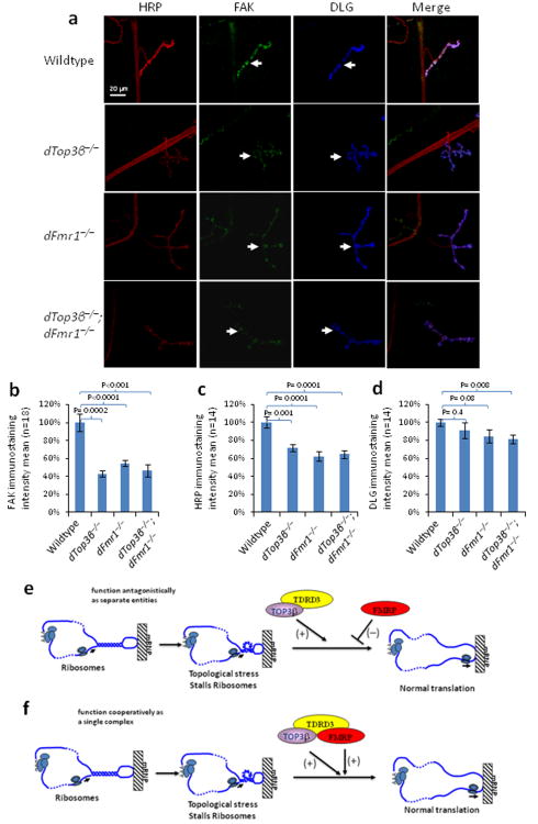
Topoisomerases are crucial for solving DNA topological problems, but they have not been linked to RNA metabolism. Here we show that human topoisomerase 3β (Top3β) is an RNA topoisomerase that biochemically and genetically interacts with FMRP, a protein that is deficient in fragile X syndrome and is known to regulate the translation of mRNAs that are important for neuronal function, abnormalities of which are linked to autism. Notably, the FMRP-Top3β interaction is abolished by a disease-associated mutation of FMRP, suggesting that Top3β may contribute to the pathogenesis of mental disorders. Top3β binds multiple mRNAs encoded by genes with neuronal functions linked to schizophrenia and autism. Expression of one such gene, that encoding protein tyrosine kinase 2 (ptk2, also known as focal adhesion kinase or FAK), is reduced in the neuromuscular junctions of Top3β mutant flies. Synapse formation is defective in Top3β mutant flies and mice, as well as in FMRP mutant flies and mice. Our findings suggest that Top3β acts as an RNA topoisomerase and works with FMRP to promote the expression of mRNAs that are crucial for neurodevelopment and mental health.


Figures

Figure 1
Figure 1. Top3β and TDRD3 form a complex that associates with FMRP; this association is disrupted by a patient-derived point mutation or by substitution of methylated arginine residues in FMRP
(a) Schematic representations of Top3α and Top3β complexes. TDRD3 and RMI1 are similar to each other in that both contain DUF1767 and OB-fold domains at their N-terminus. The unique intervening region within each OB-fold is indicated by a loop. (b-e) Silver-stained SDS gels (b,d), and immunoblotting (c,e), show the endogenous Top3β-TDRD3 complex immunoprecipitated either from whole-cell extracts by a Top3β antibody (b, c), or from nuclear extract by a TDRD3 antibody (d,e), respectively. In (d), IP was performed from nuclear extract with (+SP6) or without (-SP6) Superose 6 column fractionation (see Supplementary Fig. 1c). The major polypeptides on the gel (marked with arrows) were identified by mass spectrometry. Immunoblotting in (c,e) also shows that Top3β-TDRD3 co-immunoprecipitates with FMRP. (f) IP-Western (bottom panels) using transfected HEK293 cells shows that FMRP variants with a patient-derived I304N mutation, or the RGG-box deletion (ΔRGG), or substitution of methylarginines (mR-sub) within the RGG-box, are defective in association with Top3β-TDRD3. The mR-sub variant substituted 5 arginine residues within the RGG-box, R527K, R533K, R538K, R543K and R545H; the last 4 residues are methylated. FMRP is double-tagged with Flag and EGFP; and a Flag antibody was used for IP. A Mock IP was done using untransfected HEK293 cells. Asterisk marks a crossreactive polypeptide. A schematic representation of FMRP domain structure and various mutations was shown on top. Full-length pictures of the blots are in Supplementary Fig. 10. The representative images shown have been repeated at least twice, and the results are reproducible.
Figure 2
Figure 2. TDRD3 acts as a bridge connecting Top3β and FMRP
(a-c) IP-western to assess whether various TDRD3-deletion mutants described in (b) co-immunoprecipitate with Top3β, FMRP, FXR1 and FXR2. The Flag-tagged TDRD3 variants were transfected into HEK293 cells, and co-IP was performed using a Flag antibody. The crossreactive polypeptides are indicated with asterisks. A mock IP from untransfected HEK293 cells was included as a control. (b) Schematic representation of different TDRD3-deletion mutants (left), and their ability to coimmunoprecipitate with Top3β and FMRP from HEK293 extracts (right). (d) IP-Western shows that the association between Top3β and FMRP was disrupted in HeLa cells depleted of TDRD3 by siRNA. The IP was performed using a Flag antibody from extracts of HeLa cells stably expressing HF-Top3β. Full-length pictures of the blots are in Supplementary Fig. 10. The representative images shown have been repeated at least twice, and the results are reproducible.
Figure 3
Figure 3. Top3β associates with TDRD3 and FMRP in SGs and polyribosomes
(a) Immunofluorescence showing that GFP-Top3β transiently transfected into HeLa cells is redistributed into SGs after arsenite (As) treatment. TIA-1 is included as a SG marker. The nucleus is stained with DAPI. (b,c) Immunofluorescence analysis of GFP-Top3β co-localization with TDRD3 and FMRP in SGs, respectively. (d) Immunoblot analysis of the colocalization of Top3β and TDRD3 with polyribosomes. Cytoplasmic extracts were prepared from suspension HEK293 cells that had been treated with or without puromycin and centrifuged on a 15-60% w/w linear sucrose gradient. The direction of sedimentation of the gradient is left to right (heaviest fraction is 11). The positions of ribosomal subunits, and mono- and polyribosomes are detected by the absorption profile at 254 nM and indicated at the bottom. β-actin was included as a negative control. An asterisk marks a crossreactive polypeptide. Full-length pictures of the blots are in Supplementary Fig. 10. The representative images shown have been repeated at least twice, and the results are reproducible.
Figure 4
Figure 4. Top3β has RNA topoisomerase activity that depends on a conserved RGG RNA-binding motif
(a) Schematic representation of an RNA topoisomerase assay modified from a previous publication. A synthetic circular RNA substrate contains two pairs of complementary regions (red and green) separated by single-stranded spacers (black). Through strand passage reactions, this substrate is converted to a knot in which the two pairs of complementary regions can form normal double helices. (b) Silver-stained SDS gel showing purified recombinant wildtype or Y336F mutant HF-Top3β proteins. This mutation is known to inactivate topoisomerase activity on DNA. (c) Autoradiographs from the RNA topoisomerase assay show that Top3β, but not its catalytic mutant or Top3α, has RNA topoisomerase activity. The reaction contains 1 nM 32P-labeled circular RNA substrate and increasing concentrations of wildtype or Y336F Top3β mutant (0.05 nM, 0.1 nM, 0.2 nM, 0.4 nM, 0.8 nM or 1.6 nM), or Top3α (0.1 nM, 0.3 nM, 1 nM, 3 nM, 10 nM or 30 nM). A ladder of RNA markers and the purified RNA knot were loaded on the left of every panel. The linear breakdown products of cyclic RNA exist in all reactions. A darker exposure of the autoradiographs shows that a small amount of catenane was also generated in the reaction (Supplementary Fig. 4a). (d) Top panel: schematic representations showing that Top3β, but not Top3α, contains a RGG motif. Bottom panel: the alignment of RGG motifs of Top3β from several higher eukaryotes. The RGG boxes are indicated by underline. Arginine and Glycine are indicated by red and green letters, respectively. Conserved regions are highlighted in yellow. (e,f) Gel-shift assay (e) and its quantification (f) show that a fusion protein containing MBP and the RGG motif of Top3β (MBP-RGG) preferentially binds RNA compared to DNA. Reaction contains 1 nM single-stranded (ss) or double-stranded (ds) RNA or DNA. MBP-TDRD31-187 was included as a negative control. (g,h) RNA topoisomerase assay (g, right panel) and its quantification (h) show that the RGG motif of Top3β is important for its RNA topoisomerase activity; (g, left panel), silver-stained SDS gel of the purified wild-type or RGG motif-deleted Top3β. The representative images shown have been repeated at least twice, and the results are reproducible.
Figure 5
Figure 5. Top3β binds coding regions of mRNAs, and its bound mRNAs are enriched with match FMRP targets
(a) Autoradiograph of a SDS-PAGE gel from the HITS-CLIP assay shows that a significant amount of 32P-labeled RNA was crosslinked to HF-Top3β. A mock immunoprecipitation was performed using HeLa cells that do not express HF-Top3β. The RNA in gel slices marked H (high) and L (low) were extracted, reverse-transcribed, and sequenced; but only data from H are presented. (b) Histogram illustrates that Top3β binding sites, represented by sequence tags identified by HITS-CLIP, preferentially map to exons, but not introns. The number of sequence tags was counted at specific distances from exon start (left graph) and exon end (right graph) and then converted to tag density per 1 Kb. Data for longer exons are included in Supplementary Fig. 5. (c) A graph showing that Top3β binding sites on mRNAs are enriched in open-reading frames (ORFs), but not in 5′ or 3′ untranslated regions (UTRs). Peaks are locations with >15 tags. (d) A ranked plot shows that genes containing higher frequency of Top3β tags are enriched with FMRP targets (red line) than those with lower frequency of Top3β tags. The small inlet graph shows that the ratio between the number of FMRP targets in the top 1000 mRNAs with highest Top3β tag frequency vs. that in the bottom 1000 mRNAs with lowest Top3β tag frequency. The enrichment for HuR was included for comparison. The statistical analyses used Chi-square test. (e) A Venn diagram shows the number of top Top3β targets that overlap those of FMRP. These common targets are listed in Table S2. (f) A graph shows the top gene ontology (GO) terms that are enriched in the common targets of Top3β and FMRP. The GO terms are ranked by –log (P values). The boxes highlight the categories that may be relevant to functions of Top3β and FMRP in neurons. The genes of each category are listed in Supplementary Table 4.
Figure 6
Figure 6. Top3β and TDRD3 mutations modify dFMR1 function in Drosophila eyes
(a) Scanning Electron Microcopy (SEM) of Drosophila compound eyes shows genetic interactions of dFMR1 with Top3β and TDRD3. Different genotypes are indicated. The rough eye phenotype induced by ectopic expression of dFMR1 in the eye (sev-dFMR1) is enhanced by a Top3β null mutant, but suppressed by a reduction-of-function mutant of TDRD3(dTDRD3-P) (Supplementary Fig. 6e,f). The crater-like holes are necrotic ommatidia that were quantified and shown in (b). The magnifications are: first row, 250×; second row, 800×. The tangential sections of the eyes are shown in Supplementary Fig. 6d. (b) A graph shows the number of necrotic ommatidia in the eyes of different genotypes. For flies of each genotype, 10 eyes were counted. The p values between indicated genotypes were calculated using Student t-test and are shown on the top. The error bars represent standard errors of means (s.e.m). The representative images shown have been repeated at least twice, and the results are reproducible.
Figure 7
Figure 7. Drosophila Top3β and dFmr1 work in a common pathway to promote formation of NMJs
(a) Representative immunofluorescence images of neuromuscular junctions at muscle 4 (NMJ4) of third instar wandering Drosophila larvae of different genotypes as indicated. The NMJ4 was co-labeled with a presynaptic marker (anti-HRP, red) and a postsynaptic marker (anti-DJG, green). The same set of images is shown in both top and bottom, but the arrowheads in the top images mark synaptic boutons, and the arrows in bottom mark synaptic branches. (b) Quantification of synaptic boutons from NMJ4 segments 3 to 5 (n>=18) of different genotypes as indicated. (c) Quantification of synaptic branches at NMJ4 from segments 3, 4, and 5 of both sides of wandering third instar larvae (n>=18). The graphs show the means of the branches, and error bars represent standard errors of mean (s.e.m.). The p-values shown above each bar were calculated between wildtype (WT) and a mutant using Student' t-test. The representative images shown have been repeated at least twice, and the results are reproducible.
Figure 8
Figure 8. Drosophila Top3β and dFmr1 work in the same pathway to promote expression of ptk2/FAK
(a) Representative immunofluorescence images, and (b) their quantification, show that the level of ptk2/FAK protein (green) is reduced in NMJs of the 3rd instar larvae of Top3β and dFmr1 single and double mutant flies. Presynapses of NMJs and axons were labeled with anti-HRP antibody (Red), whereas postsynapses were labeled with anti-DLG antibody (Blue). The FAK staining of the entire NMJ area, which was marked by DLG staining [arrows in (a)], was quantified by using Imaris imaging software and shown in (b). Eighteen sets of the NMJs from wild-type or top3β mutants were stained and quantified. The p-values in the graph were calculated using Student' t-test. Error bars represent standard errors of means (s.e.m.). (c and d) Quantification of immunofluorescence signals of HRP and DLG to serve as comparisons. The representative images shown have been repeated at least twice, and the results are reproducible. (e) A model to illustrate how Top3β -TDRD3 complex may work antagonistically with FMRP to regulate translation of an mRNA. An mRNA may become topologically constrained by circularization through protein-mediated interactions between its 5′-UTR and 3′-poly A tail. The mRNA may contain local duplex regions or hairpin structures. When ribosomes or RNA helicases unwind such structures, it may create topological stress that enhances ribosomal stalling which is facilitated by FMRP binding. Top3β may reduce the topological stress and thus antagonize FMRP-mediated ribosomal stalling. Our biochemistry experiments showed that a large fraction of FMRP in cells does not associate with Top3β-TDRD3. We hypothesize that this fraction of FMRP antagonizes Top3β action. (f) A model that illustrates how Top3β -TDRD3 may cooperate with FMRP to enhance mRNA translation. Our biochemistry experiments show that a fraction of Top3β-TDRD3 complex associates with FMRP and vise versa. We hypothesize that this fraction of FMRP may enhance Top3β-TDRD3 to bind its target mRNA, reduce topological stress, and stimulate mRNA translation.

Comment in

References

    1. Wang JC. Cellular roles of DNA topoisomerases: a molecular perspective. Nat Rev Mol Cell Biol. 2002;3:430–440. - PubMed
    1. Wang H, Di Gate RJ, Seeman NC. An RNA topoisomerase. Proc Natl Acad Sci U S A. 1996;93:9477–9482. - PMC - PubMed
    1. Li W, Wang JC. Mammalian DNA topoisomerase IIIalpha is essential in early embryogenesis. Proc Natl Acad Sci U S A. 1998;95:1010–1013. - PMC - PubMed
    1. Plank JL, Chu SH, Pohlhaus JR, Wilson-Sali T, Hsieh TS. Drosophila melanogaster topoisomerase IIIalpha preferentially relaxes a positively or negatively supercoiled bubble substrate and is essential during development. J Biol Chem. 2005;280:3564–3573. - PubMed
    1. Wu J, Hou JH, Hsieh TS. A new Drosophila gene wh (wuho) with WD40 repeats is essential for spermatogenesis and has maximal expression in hub cells. Dev Biol. 2006;296:219–230. - PMC - PubMed

Publication types

MeSH terms

Substances

Associated data