. 2013:3:2360.
doi: 10.1038/srep02360.

Polypyrrole hollow microspheres as echogenic photothermal agent for ultrasound imaging guided tumor ablation

Affiliations

Polypyrrole hollow microspheres as echogenic photothermal agent for ultrasound imaging guided tumor ablation

Zhengbao Zha et al. Sci Rep. 2013.

Abstract

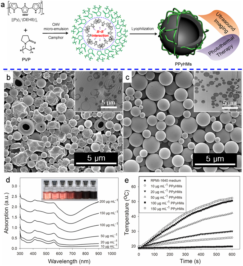
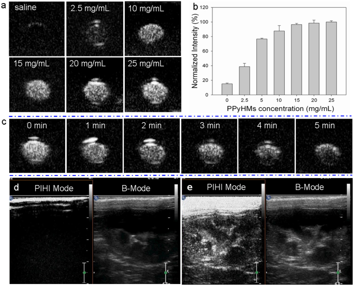
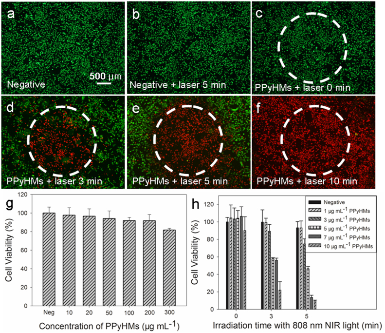
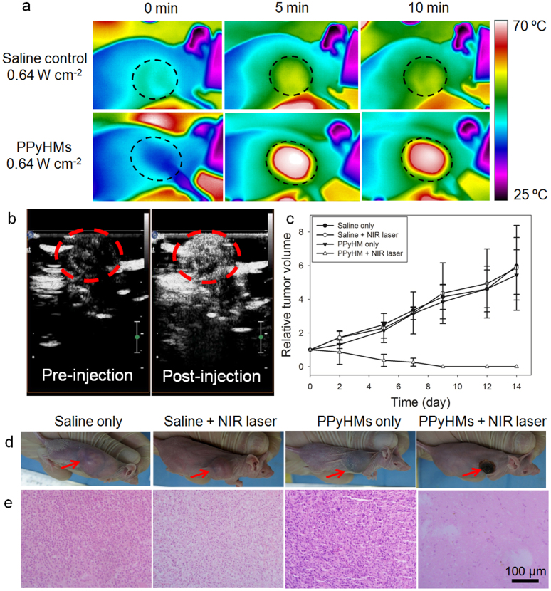
Ultrasound (US) imaging provides a valuable opportunity to administer photothermal therapy (PTT) of cancer with real-time guidance to ensure proper targeting, but only a few theranostic agents were developed by physically grafting near infrared (NIR)-absorbing inorganic nanomaterials to ready-made ultrasound contrast agents (UCAs) for US imaging guided PTT. In this paper, NIR absorbing hollow microspheres were generated from polypyrrole merely using a facile one-step microemulsion method. It was found that the obtained polypyrrole hollow microspheres (PPyHMs) can act as an efficient theranostic agent not only to enhance US imaging greatly, but also exhibit excellent photohyperthermic effects. The contrast consistently sustained the echo signals for no less than 5 min and the NIR laser light ablated the tumor completely within two weeks in the presence of PPyHMs. More importantly, no use of additional NIR absorber substantially minimizes an onetime dose of the theranostic agent.


Figures

Figure 1
Figure 1. Preparation and characterization of water-dispersible PPyHMs.
(a) Schematic illustration of the formation of echogenic PPyHMs for combined US imaging and PTT via a facile O/W microemulsion method; SEM and ultrathin-section TEM images of obtained PPyHMs with (b) PVP and (c) PVA as stabilizers; (d) UV-vis-NIR spectra of various concentrations PPyHMs dispersed in RPMI-1640 culture medium, inset was the photograph for various concentrations of PPyHMs dispersed in RPMI-1640 culture medium, indicating good dispersibility; (e) Heating curves of PPyHMs in RPMI-1640 culture medium at different concentrations under 808 nm laser irradiation.
Figure 2
Figure 2. Ultrasound imaging of PPyHMs.
In vitro ultrasound contrast-enhanced images in a latex tube (a) US images of various concentrations of PPyHMs; (b) Normalized US intensities over PPyHMs concentration; (c) Time-dependent echogenic behaviors of PPyHMs at the concentration of 20 mg mL−1. In vivo ultrasonograms in the rabbit right kidney (d) pre- and (e) post-administration of PPyHMs.
Figure 3
Figure 3. Localized photohyperthermic effect.
Photothermal destruction of U87-MG cells with or without PPyHMs and NIR laser (808 nm, 6 W cm−2) treatments (a, b, c, d, e and f). White circle indicates the laser spot; live/dead stain for viability shows dead cells as red while viable cells as green; (g) Cell viability of HUVECs with 24 h exposure to various concentrations of PPyHMs; (f) U87-MG cell viability after treatment with different concentrations of PPyHMs and NIR laser for different irradiation time.
Figure 4
Figure 4. In vivo US imaging guided photothermal therapy.
(a) IR thermal images of tumor-bearing mice with and without PPyHMs injection exposed to the NIR laser at the power density of 0.64 W cm−2 recorded at different time intervals. (b) contrast-enhanced ultrasonograms after the intratumoral injection of PPyHMs (0.2 mL, 2 mg mL−1) into the mice from PPyHMs + laser group for visualization of the agent distribution to guide the following PTT (tumors are highlighted in the red circles). (c) The tumor growth curves of different groups of mice after PTT treatment. The tumor volumes were normalized to their initial sizes. (d) Representative photographs of mice bearing U87-MG tumors after various different treatments indicated. (e) H&E stained tumor slices collected from different groups of mice immediately after laser irradiation. The PPyHMs injected tumor was severely damaged after laser irradiation.

References

    1. Vogel A. & Venugopalan V. Mechanisms of pulsed laser ablation of biological tissues. Chem. Rev. 103, 577–644 (2003). - PubMed
    1. Fiedler V. U. et al. Laser-induced interstitial thermotherapy of liver metastases in an interventional 0.5 tesla MRI system: Technique and first clinical experiences. J. Magn. Reson. Imaging 13, 729–737 (2001). - PubMed
    1. Shao J. et al. Photothermal nanodrugs: potential of TNF-gold nanospheres for cancer theranostics. Sci. Rep. 3, 1293 (2013). - PMC - PubMed
    1. Yang K. et al. Multimodal imaging guided photothermal therapy using functionalized graphene nanosheets anchored with magnetic nanoparticles. Adv. Mater. 24, 1868–1872 (2012). - PubMed
    1. Yu M. K. et al. Image-guided prostate cancer therapy using aptamer-functionalized thermally cross-linked superparamagnetic iron oxide nanoparticles. Small 7, 2241–2249 (2011). - PubMed

Publication types

LinkOut - more resources