. 2013 Sep;10(9):896-902.
doi: 10.1038/nmeth.2592. Epub 2013 Aug 4.

Automated identification of functional dynamic contact networks from X-ray crystallography

Affiliations

Automated identification of functional dynamic contact networks from X-ray crystallography

Henry van den Bedem et al. Nat Methods. 2013 Sep.

Abstract

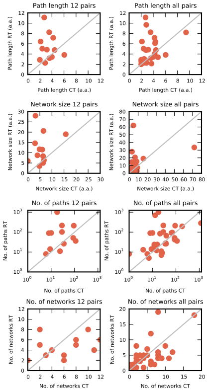
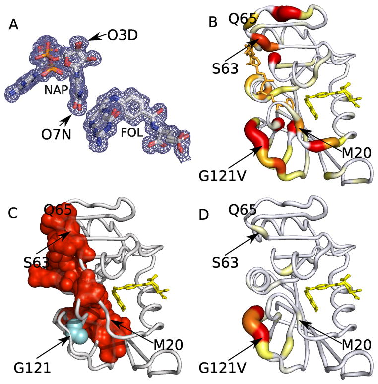
Protein function often depends on the exchange between conformational substates. Allosteric ligand binding or distal mutations can stabilize specific active-site conformations and consequently alter protein function. Observing alternative conformations at low levels of electron density, in addition to comparison of independently determined X-ray crystal structures, can provide mechanistic insights into conformational dynamics. Here we report a new algorithm, CONTACT, that identifies contact networks of conformationally heterogeneous residues directly from high-resolution X-ray crystallography data. Contact networks determined for Escherichia coli dihydrofolate reductase (ecDHFR) predict the observed long-range pattern of NMR chemical shift perturbations of an allosteric mutation. A comparison of contact networks in wild-type and mutant ecDHFR suggests that mutations that alter optimized contact networks of coordinated motions can impair catalytic function. CONTACT-guided mutagenesis can exploit the structure-dynamics-function relationship in protein engineering and design.


Conflict of interest statement

The authors declare no competing financial interests.

Figures

Figure 1
Figure 1
Mechanisms for conformational exchange in Cyclophilin A (a) X-ray electron density map contoured at 1 σ (blue mesh) and 0.3 σ (cyan mesh) of CYPA is fit with discrete alternative conformations using qFit. Alternative conformations are colored red, orange, or yellow, with hydrogen atoms added in green. (b) Visualizing a pathway in CYPA: atoms involved in clashes are shown in spheres scaled to van der Waals radii and clashes between atoms highlighted by cyan dashes. This pathway originates with the OG atom of Ser99 conformation A and the CE1 atom of Phe113 conformation B, which clash to 0.8 of their summed van der Waals radii. The pathway progresses from Phe113 to Gln63 and after moving Met61 to conformation B introduces no new clashes the pathway is terminated. A 90 degree rotation of the final panel is shown to highlight how the final move of Met61 relieves the clash with Gln63. (c) Networks identified by CONTACT are displayed as nodes connected by edges representing contacts that clash and are relieved by alternative conformations. The pathway in b forms part of the red contact network in CYPA and is highlighted by the dark purple edges. (d) The six contact networks comprising 29% of residues are mapped on the three dimensional structure of CYPA. The contact network shown in red overlaps with the dynamic network identified by NMR chemical shift perturbation and relaxation dispersion experiments.
Figure 2
Figure 2
Characteristics of pathways and contact networks are sensitive to temperature. Changes to average pathway lengths, contact network sizes, number of pathways, and number of contact networks across 12 closely matched high resolution (left column) or all matched (right column) room temperature (RT) and cryogenic datasets (CT). Each data point represents paired data sets, with values corresponding to room temperature along the vertical axis, and cryogenic temperature along the horizontal axis. Data points are expected to lie along a 45° degree line if there were no differences between the room and cryogenic temperature pairs.
Figure 3
Figure 3
All-atom contact networks in ecDHFR. (a) Contact networks are displayed in surface rendering on the crystal structure of the room temperature E:NADP+:FOL complex (3QL3) (top panels) above the largest contact network graphs (bottom panels). Nine all-atom contact networks comprise 47% of the residues in the room temperature model of ecDHFR. The NADP+ cofactor is part of the red contact network, and shown in red spheres. Folate, part of the yellow contact network, is shown in yellow spheres. The red network connects residue Phe125 in the FG loop to the adenosine binding domain (Ser63–Gln65) exclusively through the NADP cofactor. Residues in the cyan (b) and green (c) contact networks broadly agree with those identified undergoing collective exchanges in CPMG relaxation dispersion experiments in a process that is distinct from the conformational exchange observed near the active site. Consistent with NMR data, we observed that the cyan and green contact networks do not contact active site residues. The salmon and blue contact networks (c) are implicated in hinge motions. The orange contact network is implicated in changing hydrogen bonding patterns during the closed to occluded transition of the M20 loop. The yellow contact network links active site residues I5 and I95 to the folate.
Figure 4
Figure 4
Contact network analysis and an allosteric mutant (G121V) of ecDHFR. (a) A 2mFo-DFc electron density map around the cofactor and substrate of the room temperature (E:NADP+:FOL) complex contoured at 0.3 σ. Asymmetric density profiles around oxygen atoms (O7N, O3D) of the NADP molecule support multiple conformations. (b) The color (yellow to red) and thickness of the backbone tube represents the magnitude of the weighted chemical shift differences obtained from WT and a G121V mutant (E:NADP+:FOL) complex from 0.1–1ppm. Residues Ser63, Gln65, Gly67, and Thr68 in the adenosine binding domain exhibit large chemical shift changes despite their location over 23 Å away from the mutation site. The G121V mutation stabilizes the occluded conformation of the enzyme. In the WT E:NADP+:FOL complex, the M20 loop is in the “closed” conformation, whereas in the corresponding complex of G121V the M20 loop is in the “occluded” conformation. (c) The red contact network obtained from the room temperature WT (E:NADP+:FOL) complex (also Fig. 3a) in the same orientation. For illustrative purposes the G121V mutation (cyan) is modeled on the WT molecule, abutting the red contact network. Long-range conformational coupling generally corresponds to chemical shift propagation, while local effects owing to increased flexibility of the FG loop in the mutant complex are absent. (d) Chemical shift differences between binary WT and G121V mutant (E:FOL) complexes are localized to the site of mutation, confirming the central role of the NADP cofactor in coupling distant sites in ecDHFR.
Figure 5
Figure 5
Increased conformational heterogeneity at the active site of the crystal structure of the E:NADP+:FOL complex of the N23PP/S148A mutant. (a) The red contact network shares similarities with its WT counterpart, but several residues in the Met20 loop now participate in this contact network. The connection between the FG loop and NAP to the adenosine binding domain through residue I14 has been replaced by Met20 loop residues Met20, Pro21, and Trp22. (b) The cyan contact network is largely comprised of three beta strands towards the C terminus of the molecule. (c) An isomorphous difference electron density map of the WT and N23PP/S148A datasets phased with the WT crystal structure. Positive difference density (4.0σ green) in the absence of negative difference density (−4.0σ red) indicates elevated heterogeneity. Mutation sites are shown with spheres, NADP+ in orange and folate in yellow (see also Supplementary Figure 7b).

Comment in

References

    1. Fraser JS, Jackson CJ. Mining electron density for functionally relevant protein polysterism in crystal structures. Cell Mol Life Sci. 2011;68:1829–1841. - PMC - PubMed
    1. Karplus M, Kuriyan J. Molecular dynamics and protein function. Proc Natl Acad Sci USA. 2005;102:6679–85. - PMC - PubMed
    1. Marlow MS, Dogan J, Frederick KK, Valentine KG, Wand AJ. The role of conformational entropy in molecular recognition by calmodulin. Nat Chem Biol. 2010;6:352–8. - PMC - PubMed
    1. Fraser JS, et al. Hidden alternative structures of proline isomerase essential for catalysis. Nature. 2009;462:669–73. - PMC - PubMed
    1. Yu B, et al. Structural and energetic mechanisms of cooperative autoinhibition and activation of Vav1. Cell. 2010;140:246–56. - PMC - PubMed

Publication types

MeSH terms