. 2013 Sep 12;501(7466):252-6.
doi: 10.1038/nature12428. Epub 2013 Aug 4.

Stability and function of regulatory T cells is maintained by a neuropilin-1-semaphorin-4a axis

Affiliations

Stability and function of regulatory T cells is maintained by a neuropilin-1-semaphorin-4a axis

Greg M Delgoffe et al. Nature. .

Abstract

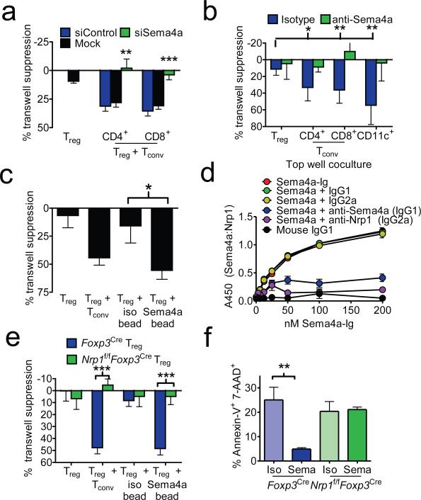
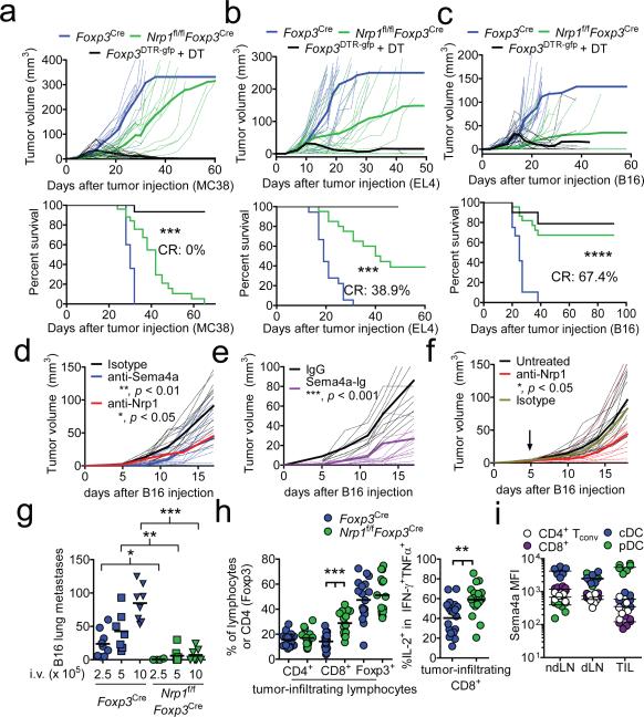
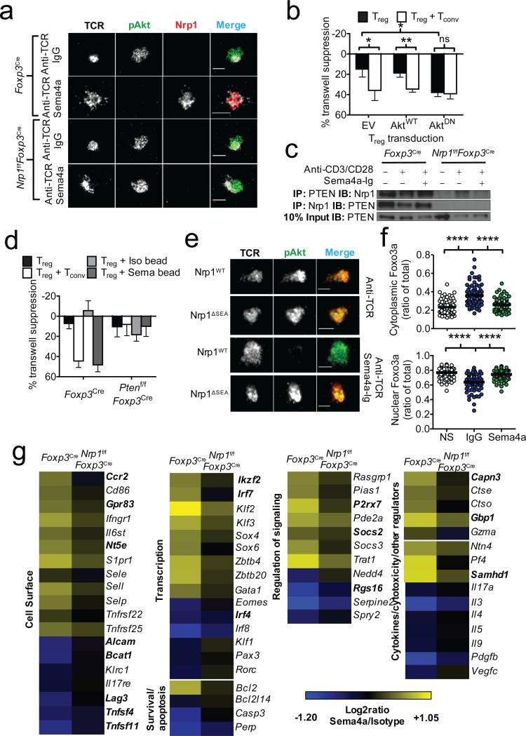
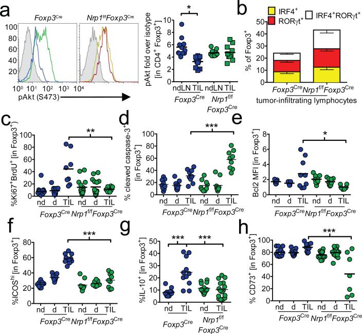
Regulatory T cells (Treg cells) have a crucial role in the immune system by preventing autoimmunity, limiting immunopathology, and maintaining immune homeostasis. However, they also represent a major barrier to effective anti-tumour immunity and sterilizing immunity to chronic viral infections. The transcription factor Foxp3 has a major role in the development and programming of Treg cells. The relative stability of Treg cells at inflammatory disease sites has been a highly contentious subject. There is considerable interest in identifying pathways that control the stability of Treg cells as many immune-mediated diseases are characterized by either exacerbated or limited Treg-cell function. Here we show that the immune-cell-expressed ligand semaphorin-4a (Sema4a) and the Treg-cell-expressed receptor neuropilin-1 (Nrp1) interact both in vitro, to potentiate Treg-cell function and survival, and in vivo, at inflammatory sites. Using mice with a Treg-cell-restricted deletion of Nrp1, we show that Nrp1 is dispensable for suppression of autoimmunity and maintenance of immune homeostasis, but is required by Treg cells to limit anti-tumour immune responses and to cure established inflammatory colitis. Sema4a ligation of Nrp1 restrained Akt phosphorylation cellularly and at the immunologic synapse by phosphatase and tensin homologue (PTEN), which increased nuclear localization of the transcription factor Foxo3a. The Nrp1-induced transcriptome promoted Treg-cell stability by enhancing quiescence and survival factors while inhibiting programs that promote differentiation. Importantly, this Nrp1-dependent molecular program is evident in intra-tumoral Treg cells. Our data support a model in which Treg-cell stability can be subverted in certain inflammatory sites, but is maintained by a Sema4a-Nrp1 axis, highlighting this pathway as a potential therapeutic target that could limit Treg-cell-mediated tumour-induced tolerance without inducing autoimmunity.


Figures

Figure 1
Figure 1. Sema4a binds Nrp1 to potentiate Treg function and survival in vitro
a, Transwell suppression assay in which Treg cells were cocultured in the top chamber of a transwell plate with anti-CD3/anti-CD28 coated beads in the presence or absence of CD4+ or CD8+ Tconv cells that had been previously transfected with scrambled or siRNA to Sema4a. Proliferation of Tconv cells stimulated with anti-CD3/anti-CD28 coated beads in the bottom chambers was measured by [3H]-thymidine uptake. b, Transwell suppression assay with Treg cells cocultured in top chamber with CD4+, CD8+, or CD11c+ cells including anti-Sema4a or its isotype control. c, Transwell suppression assay in which Treg were cocultured in the absence of Tconv but in the presence of beads coated with Sema4a-Ig or its isotype control. d, ELISA based binding assay in which plates coated with Nrp1 were incubated with Sema4a-Ig or IgG1 in the presence of various blocking antibodies. Sema4a-Ig was detected using an isotype specific antibody. e, Transwell suppression assay where Treg were purified by flow cytometry from Foxp3Cre or Nrp1f/fFoxp3Cre mice. f, Annexin V-7-AAD staining of Treg stimulated for 48 h in vitro in the presence of Sema4a-Ig or its isotype control. Results represent the mean of five independent experiments. *, p < 0.05, **, p < 0.01, ***, p < 0.001 by unpaired t-test. Error bars indicate s.e.m.
Figure 2
Figure 2. Nrp1-deficient Treg fail to suppress antitumor immune responses
a, Tumor growth curve (top) and survival plot (bottom) of Foxp3Cre, Nrp1f/fFoxp3Cre, or Foxp3DTR.gfp mice receiving 1.25 × 10⁵ MC38 melanoma cells s.c. and (for Foxp3DTR.gfp) 100 μg diphtheria toxin (DT) i.p. twice weekly. b, As in a, but mice received 1.25 × 10⁵ EL4 thymoma i.d. c, As in a, but mice received 1.25 × 10⁵ B16 melanoma i.d. d, Tumor growth curve of C57/BL6 mice receiving 1.25 × 10⁵ B16 melanoma i.d. concomitant with injections of isotype control, anti-Sema4a, or anti-Nrp1 (100 μg) twice weekly. e, Tumor growth curve as in d except mice received Sema4a-Ig twice weekly. f, Tumor growth curve of C57/BL6 mice receiving 1.25 × 10⁵ B16 melanoma i.d. When tumors were palpable (day 5, indicated by arrow), mice began receiving injections of anti-Nrp1 or its isotype control (400 μg initially, 200 μg every 3 d). g, Lung metastasis counts from Foxp3Cre or Nrp1f/fFoxp3Cre mice injected with 2.5-10 × 105 B16 cells i.v. 17-20 d earlier. h, Tabulation of flow cytometric analysis of tumor-infiltrating lymphocytes from Foxp3Cre or Nrp1f/fFoxp3Cre mice injected i.d. with B16 18 d earlier. i, Sema4a expression of various immune cells in ndLN, dLN, or TIL. Results represent the mean of five (a-c, n=10-25 mice), three (d-h n=8-20 mice), or three (i) experiments. *, p < 0.05, **, p < 0.01, ***, p < 0.001, by (a-f) one-way ANOVA or (g-i) unpaired t-test. Error bars indicate s.e.m.
Figure 3
Figure 3. Ligation of Nrp1 by Sema4a promotes Treg stability through modulation of Akt-mTOR signaling
a, TIRF microscopic analysis of IS Akt phosphorylation in Tregs stimulated 20 min on a lipid bilayer coated with anti-TCR antibodies in the presence or absence of Sema4a-Ig. b, Transwell suppression assay using Tregs retrovirally expressing WT or DN-Akt. Transductants were selected and expanded using puromycin and IL-2. c, Immunoprecipitation (IP) analysis of Nrp1 using in vitro expanded Tregs serum starved for 3h, then stimulated as indicated for 6 h prior to IP. d, Transwell suppression assay using Foxp3Cre or Ptenf/fFoxp3Cre Tregs. e, TIRF microscopic analysis of IS Akt phosphorylation as in a of Nrp1f/fFoxp3Cre Treg cells retrovirally reconstituted with WT or ΔSEA Nrp1. f, Foxo3a cytoplasmic (top) and nuclear (bottom) localization signals, as defined by masking using actin and DNA staining. Arbitrary Units (AU) represent fluorescence intensity calculated volumetrically through 20-30 slices of Treg cells. n=70-93 g, Heat map (right) of genes regulated by Nrp1. Foxp3Cre and Nrp1f/fFoxp3Cre CD45RBlo Foxp3 (YFP)+ CD4+ T cells were stimulated for 48h with anti-CD3, anti-CD28, 100 U/mL rhIL-2, and immobilized IgG1 or Sema4a-Ig. RNA was subjected to Affymetrix gene profiling. Treg signature genes are in bold. All genes shown met FDR < 0.10 and compared using two-way ANOVA. Results represent at least three independent experiments (a, c, e, f) or represent means of three (b, d) or seven (g) experiments. *, p < 0.05, ** p < 0.01 by unpaired t-test. Error bars indicate s.e.m.
Figure 4
Figure 4. Tumor-infiltrating Treg bear a signature similar to Sema4a:Nrp1 ligation
a, Akt phosphorylation in Treg. Tumor-bearing Foxp3Cre or Nrp1f/fFoxp3Cre mice were sacrificed and ndLN and TIL were harvested. Cells were immediately fixed and stained for phospho-Akt. Shaded histogram indicates isotype control. Results are tabulated normalized to isotype control staining. IRF4/RORγt (b), Ki67/BrdU (c), cleaved caspase-3 (d), Bcl2 (e), ICOS (f), IL-10 (g) and CD73 (h) staining from ndLN, dLN, or TIL from tumor-bearing Foxp3Cre or Nrp1f/fFoxp3Cre mice. Ki67/BrdU analysis included injection with BrdU 14 h prior to harvest. IL-10 staining included restimulation with PMA and ionomycin for 16h in the presence of brefeldin A. Results represent the mean of 3-5 independent experiments. * p < 0.05, ** p < 0.01, *** p < 0.001 by paired t-test (a, n=7) or unpaired t-test (b-h n=9-25). Error bars indicate s.e.m.

References

    1. Vignali DA, Collison LW, Workman CJ. How regulatory T cells work. Nat Rev Immunol. 2008;8:523–532. - PMC - PubMed
    1. Fontenot JD, Gavin MA, Rudensky AY. Foxp3 programs the development and function of CD4+CD25+ regulatory T cells. Nat Immunol. 2003;4:330–336. - PubMed
    1. Hori S, Nomura T, Sakaguchi S. Control of regulatory T cell development by the transcription factor Foxp3. Science. 2003;299:1057–1061. - PubMed
    1. Miyao T, et al. Plasticity of Foxp3(+) T cells reflects promiscuous Foxp3 expression in conventional T cells but not reprogramming of regulatory T cells. Immunity. 2012;36:262–275. - PubMed
    1. Rubtsov YP, et al. Stability of the regulatory T cell lineage in vivo. Science. 2010;329:1667–1671. - PMC - PubMed

Publication types

MeSH terms

Associated data