

Natural history and prognostic value of corticospinal tract Wallerian degeneration in intracerebral hemorrhage
- PMID: 23913508
- PMCID: PMC3828779
- DOI: 10.1161/JAHA.113.000090
Natural history and prognostic value of corticospinal tract Wallerian degeneration in intracerebral hemorrhage
Abstract
Background: The purpose of this study was to define the incidence, imaging characteristics, natural history, and prognostic implication of corticospinal tract Wallerian degeneration (CST-WD) in spontaneous intracerebral hemorrhage (ICH) using serial MR imaging.
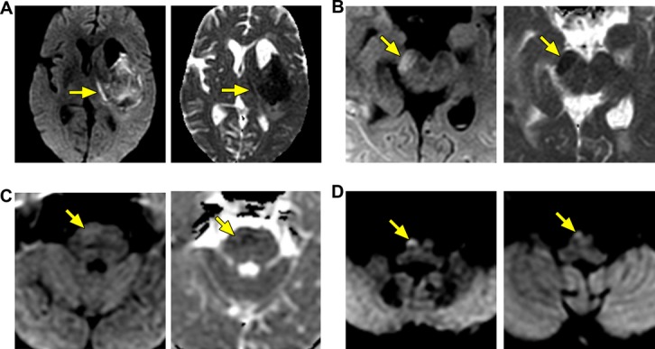
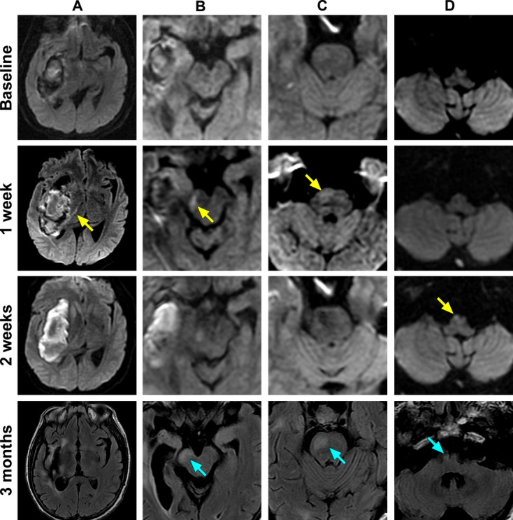
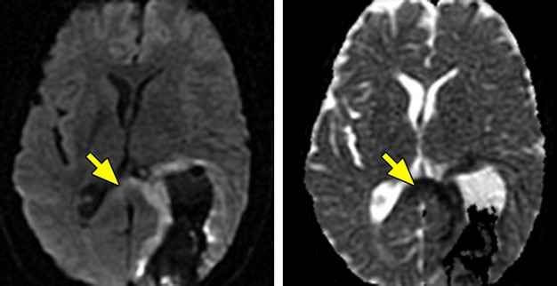
Methods and results: Consecutive ICH patients with supratentorial ICH prospectively underwent serial MRIs at 2, 7, 14, and 21 days. MRIs were analyzed by independent raters for the presence and topographical distribution of CST-WD on diffusion-weighted imaging (DWI). Baseline demographics, hematoma characteristics, ICH score, and admission National Institute of Health Stroke Score (NIHSS) were systematically recorded. Functional outcome at 3 months was assessed by the modified Rankin Scale (mRS) and the motor-NIHSS. Twenty-seven patients underwent 93 MRIs; 88 of these were serially obtained in the first month. In 13 patients (48%), all with deep ICH, CST-WD changes were observed after a median of 7 days (interquartile range, 7 to 8) as reduced diffusion on DWI and progressed rostrocaudally along the CST. CST-WD changes evolved into T2-hyperintense areas after a median of 11 days (interquartile range, 6 to 14) and became atrophic on MRIs obtained after 3 months. In univariate analyses, the presence of CST-WD was associated with poor functional outcome (ie, mRS 4 to 6; P=0.046) and worse motor-NIHSS (5 versus 1, P=0.001) at 3 months.
Conclusions: Wallerian degeneration along the CST is common in spontaneous supratentorial ICH, particularly in deep ICH. It can be detected 1 week after ICH on DWI and progresses rostrocaudally along the CST over time. The presence of CST-WD is associated with poor motor and functional recovery after ICH.
Keywords: diffusion‐weighted imaging; intracerebral hemorrhage; magnetic resonance imaging; natural history; prognosis; wallerian degeneration.
Figures
References
-
- Waller A. Experiments on the section of the glossopharyngeal and hypoglossal nerves of the frog and observations of the alterations produced thereby in the structure of their primitive fibre. Philos Trans R Soc. 1850; 140:423-429
-
- Sawlani V, Gupta R, Singh M, Kohli A. MRI demonstration of wallerian degeneration in various intracranial lesions and its clinical implications. J Neurol Sci. 1997; 146:103-108 - PubMed
-
- Ajilogba K, Rao P. Diffusion‐weighted imaging of wallerian degeneration in non‐accidental head injury. Pediatr Radiol. 2006; 36:1326. - PubMed
-
- Cobb S, Mehringer C. Wallerian degeneration in a patient with schilder disease: MR imaging demonstration. Radiology. 1987; 162:521-522 - PubMed
-
- Orita T, Tsurutani T, Izumihara A, Kajiwara K. Early, evolving wallerian degeneration of the pyramidal tract in cerebrovascular diseases: MR study. J Comput Assist Tomogr. 1994; 18:943-946 - PubMed
Publication types
MeSH terms
Supplementary concepts
Grants and funding
LinkOut - more resources
Full Text Sources
Other Literature Sources
Molecular Biology Databases
