. 2013 Sep 13;288(37):26810-9.
doi: 10.1074/jbc.M113.497032. Epub 2013 Aug 2.

The linker region plays a regulatory role in assembly and activity of the Vps4 AAA ATPase

Affiliations

The linker region plays a regulatory role in assembly and activity of the Vps4 AAA ATPase

Anna Shestakova et al. J Biol Chem. .

Abstract

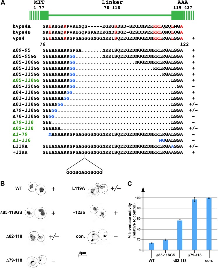
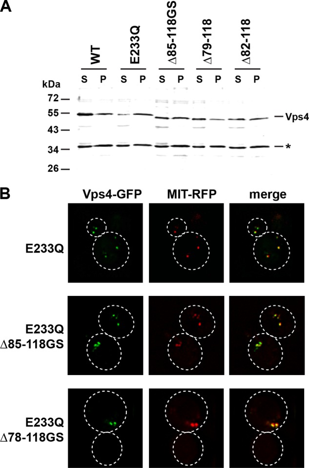
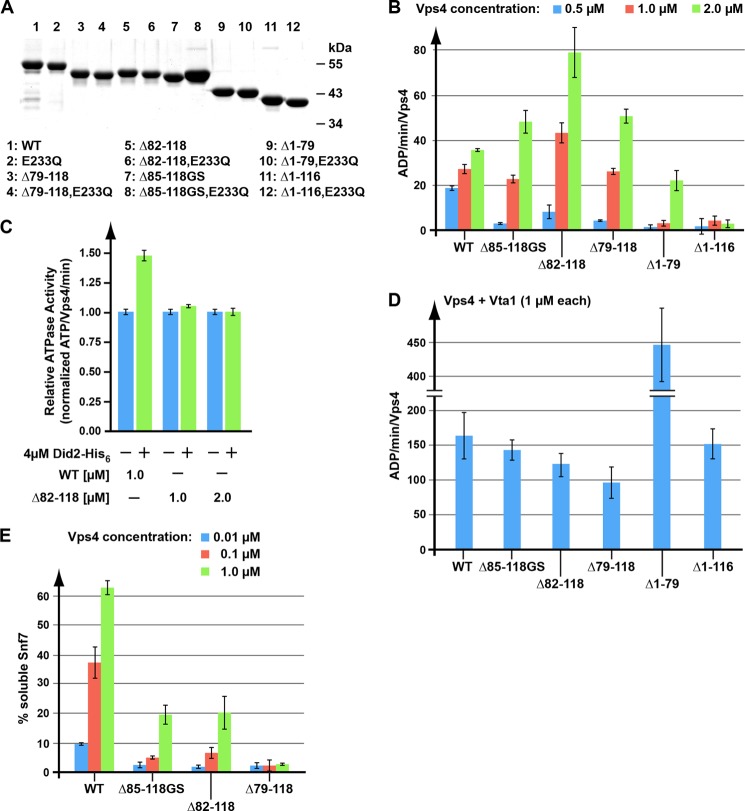
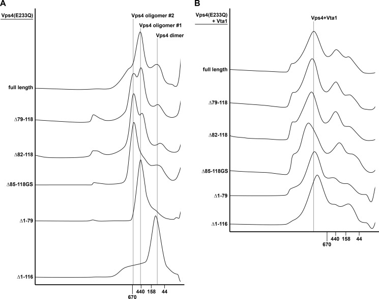
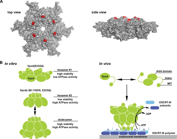
The AAA-type ATPase Vps4 functions with components of the ESCRT (endosomal sorting complex required for transport) machinery in membrane fission events that are essential for endosomal maturation, cytokinesis, and the formation of retroviruses. A key step in these events is the assembly of monomeric Vps4 into the active ATPase complex, which is aided in part by binding of Vps4 via its N-terminal MIT (microtubule interacting and trafficking) domain to its substrate ESCRT-III. We found that the 40-amino acid linker region between the MIT and the ATPase domain of Vps4 is not required for proper function but plays a role in regulating Vps4 assembly and ATPase activity. Deletion of the linker is expected to bring the MIT domains into close proximity to the central pore of the Vps4 complex. We propose that this localization of the MIT domain in linker-deleted Vps4 mimics a repositioning of the MIT domain normally caused by binding of Vps4 to ESCRT-III. This structure would allow the Vps4 complex to engage ESCRT-III subunits with both the pore and the MIT domain simultaneously, which might be essential for the ATP-driven disassembly of ESCRT-III.

Keywords: ATPases; Endosomes; Enzyme Mechanisms; Membrane; Yeast.


Figures

FIGURE 1.
FIGURE 1.
Trafficking phenotypes of vps4 linker mutants. A, amino acid sequence alignment of the linker regions of yeast and human (h) Vps4 proteins. The linker mutations analyzed in this study are listed. Dotted regions indicate deletions, whereas amino acid exchanges are labeled in blue. The mutants important for this study are highlighted in green. The +, +/−, and − labels indicate the MVB sorting efficiency of GFP-Cps1 in the corresponding vps4 mutant background. B, fluorescence microscopy of wild-type and vps4 mutants expressing GFP-Cps1. The images are inverted for better visualization of the GFP localization. The +, +/−, and − labels refer to the GFP-Cps1 sorting efficiency. con., control. C, CPY-Invertase secretion assays. The invertase activity is shown relative to the negative control vps4Δ (100%). Error bars indicate S.D.
FIGURE 2.
FIGURE 2.
Subcellular localization of mutant Vps4 proteins. A, Western blot analysis of soluble (S) and membrane-bound pellet (P) cell fractions using anti-Vps4 antiserum. The cross-reacting protein at 35 kDa (*) serves as a loading control. B, fluorescence microscopy of cells expressing GFP-tagged versions of Vps4 mutants and the MVB marker MIT-RFP.
FIGURE 3.
FIGURE 3.
In vitro analysis of linker-deleted Vps4 proteins. A, SDS-PAGE analysis of the recombinant Vps4 proteins used in the in vitro assays. Proteins are visualized by Coomassie Blue staining. B–D, Vps4 ATPase activity in the absence (B) or presence of the substrate Did2 (C) or the co-factor Vta1 (D). E, in vitro ESCRT-III disassembly by wild-type and mutant Vps4. Error bars in panels B–E indicate S.D.
FIGURE 4.
FIGURE 4.
Gel filtration analysis of wild-type or mutant Vps4 in the presence (B) or absence (A) of Vta1. All experiments were performed in the presence of 1 mm ATP.
FIGURE 5.
FIGURE 5.
Analysis of the quaternary structure of Vps4. A, recombinant Vps4 protein was cross-linked by glutaraldehyde in the presence of ADP or ATP, and the products were analyzed by SDS-PAGE (4–15% gradient gel) followed by Coomassie Blue staining. The GF lanes contain the cross-linking products of the Vps4 peak fractions from the gel filtration analyses shown in Fig. 4A. B, analytical ultracentrifugation of Vps4(Δ85–118GS, E233Q) and Vps4(E233Q) in the presence of ATP. The interference signal from scans taken at equilibrium at 5000 rpm is plotted versus the distance from the axis of rotation (radius) as red, with open symbols representing different protein concentrations (squares, 3 mg/ml; circles, 1.5 mg/ml; diamonds, 0.75 mg/ml). The black solid line represents a global fit over all concentrations and including data taken at 3000 rpm (not shown). Residuals are shown below for all concentrations.
FIGURE 6.
FIGURE 6.
Model of the MIT-regulated assembly of Vps4. A, model for the hexameric structure of the Vps4 AAA domain based on the p97 D1 structure (6). The first amino acid of the AAA domain (Leu-119) is labeled in red. B, model for the oligomerization of Vps4 into a double-ring structure triggered by either linker deletion (in vitro) or substrate binding (in vivo).

References

    1. Hill C. P., Babst M. (2012) Structure and function of the membrane deformation AAA ATPase Vps4. Biochim. Biophys. Acta 1823, 172–181 - PMC - PubMed
    1. McCullough J., Colf L. A., Sundquist W. I. (2013) Membrane fission reactions of the mammalian ESCRT pathway. Annu. Rev. Biochem. 82, 663–692 - PMC - PubMed
    1. White S. R., Lauring B. (2007) AAA+ ATPases: achieving diversity of function with conserved machinery. Traffic 8, 1657–1667 - PubMed
    1. Wendler P., Ciniawsky S., Kock M., Kube S. (2012) Structure and function of the AAA+ nucleotide binding pocket. Biochim. Biophys. Acta 1823, 2–14 - PubMed
    1. Babst M., Wendland B., Estepa E. J., Emr S. D. (1998) The Vps4p AAA ATPase regulates membrane association of a Vps protein complex required for normal endosome function. EMBO J. 17, 2982–2993 - PMC - PubMed

Publication types

MeSH terms

Substances

LinkOut - more resources