. 2013 Oct 1;305(7):H960-8.
doi: 10.1152/ajpheart.00858.2012. Epub 2013 Aug 2.

Mitochondrial reactive oxygen species: which ROS signals cardioprotection?

Affiliations

Mitochondrial reactive oxygen species: which ROS signals cardioprotection?

Anders O Garlid et al. Am J Physiol Heart Circ Physiol. .

Abstract

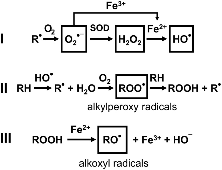
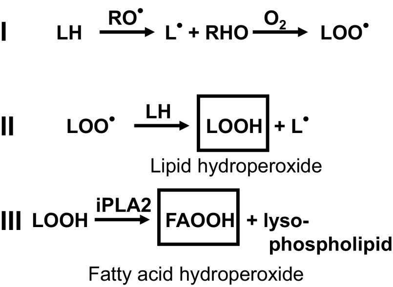
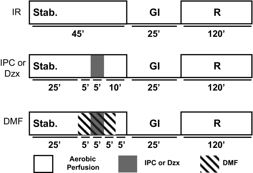
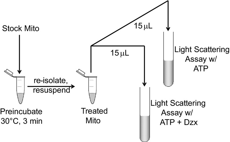
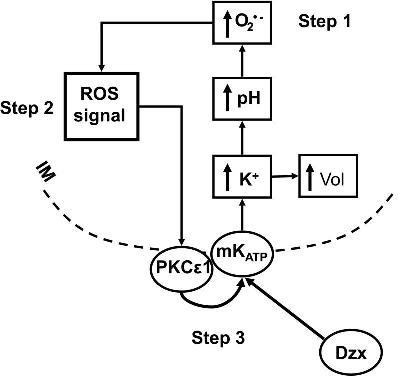
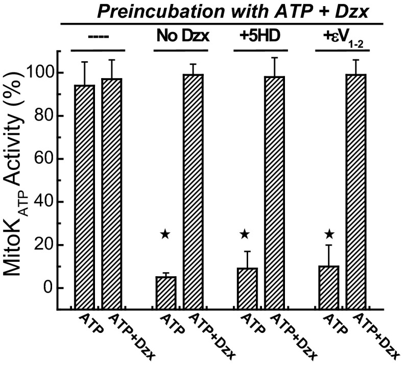
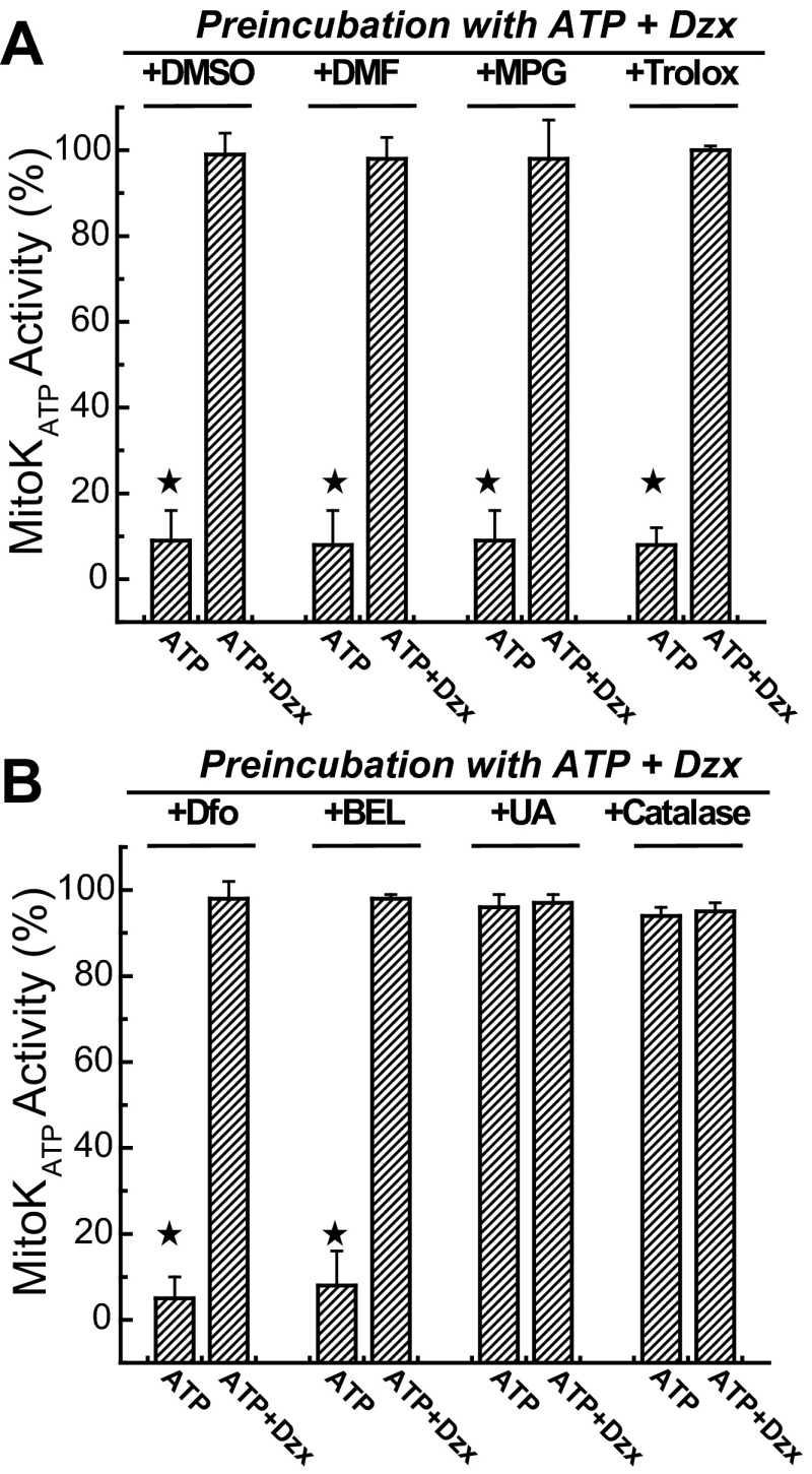
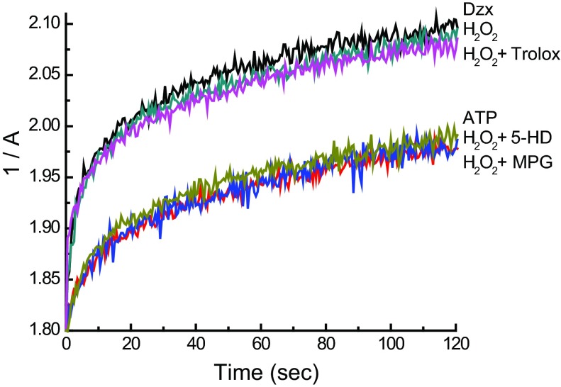
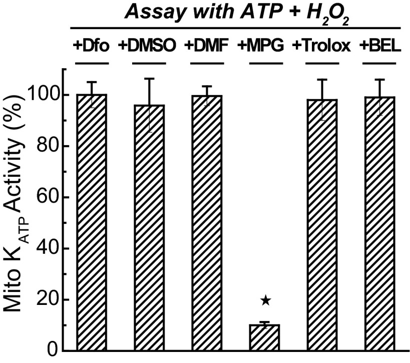
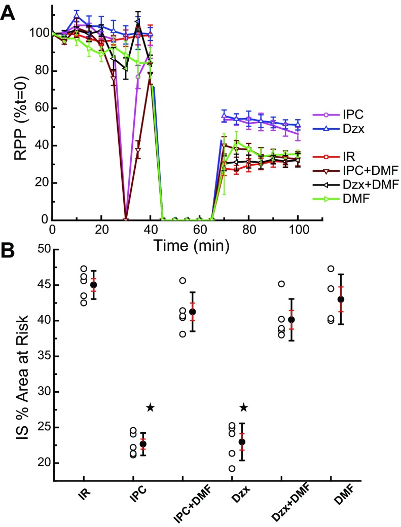
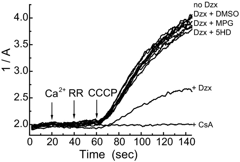
Mitochondria are the major effectors of cardioprotection by procedures that open the mitochondrial ATP-sensitive potassium channel (mitoKATP), including ischemic and pharmacological preconditioning. MitoKATP opening leads to increased reactive oxygen species (ROS), which then activate a mitoKATP-associated PKCε, which phosphorylates mitoKATP and leaves it in a persistent open state (Costa AD, Garlid KD. Am J Physiol Heart Circ Physiol 295, H874-H882, 2008). The ROS responsible for this effect is not known. The present study focuses on superoxide (O2(·-)), hydrogen peroxide (H2O2), and hydroxyl radical (HO(·)), each of which has been proposed as the signaling ROS. Feedback activation of mitoKATP provides an ideal setting for studying endogenous ROS signaling. Respiring rat heart mitochondria were preincubated with ATP and diazoxide, together with an agent being tested for interference with this process, either by scavenging ROS or by blocking ROS transformations. The mitochondria were then assayed to determine whether or not the persistent phosphorylated open state was achieved. Dimethylsulfoxide (DMSO), dimethylformamide (DMF), deferoxamine, Trolox, and bromoenol lactone each interfered with formation of the ROS-dependent open state. Catalase did not interfere with this step. We also found that DMF blocked cardioprotection by both ischemic preconditioning and diazoxide. The lack of a catalase effect and the inhibitory effects of agents acting downstream of HO(·) excludes H2O2 as the endogenous signaling ROS. Taken together, the results support the conclusion that the ROS message is carried by a downstream product of HO(·) and that it is probably a product of phospholipid oxidation.

Keywords: KATP channels; ROS signaling; cardiac ischemia; cardioprotection; mitochondria; reactive oxygen species.


Figures

Fig. 1.
Fig. 1.
Mitochondrial electron transport chain-derived ROS and formation of alkyl peroxyl alkoxyl radicals. I: superoxide (O2·−) is formed by single electron transfer to molecular oxygen (O2) from a reduced group (Ṙ) in the respiratory chain and converted to hydrogen peroxide (H2O2) by superoxide dismutase (SOD). Hydroxyl radical (HO˙) is formed from H2O2 in the presence of a transition metal ion such as iron (Fe2+/3+; the Fenton reaction). II: subsequent reactions include the formation of alkyl peroxyl radicals (ROȮ) and alkyl hydroperoxides (ROOH). HO˙ acts on an alkyl side chain (RH) to form a reduced group (Ṙ) that leads to the formation of ROȮ, and these react with RH to produce ROOH. III: alkoxyl radicals (RȮ) are formed in the decomposition of hydroperoxides (ROOH) by transition metal ions (Fe2+).
Fig. 2.
Fig. 2.
Lipid peroxidation and release of hydroperoxy fatty acids. I and II: alkoxyl (RȮ), hydroxyl (RHO), and alkyl peroxyl radicals (LOȮ) can initiate the nonenzymatic chain reaction leading to lipid peroxidation (LOOH). III: lipid hydroperoxides (LOOH) can be released by the action of mitochondrial phospholipases, such as Ca2+-independent phospholipases PLA2 (iPLA2), which have been found to modulate the function of mitochondria [see Cedars et al. (8) for a review]. The action by iPLA2 results in a hydroperoxy fatty acid (FAOOH).
Fig. 3.
Fig. 3.
Isolated heart protocols. Hearts were perfused with buffer, as described in research design and methods. After 45 min of stabilization, hearts were subjected to 25 min global ischemia (GI), followed by 2 h of reperfusion (R) and, finally, processing for infarct size estimation. Protocol for ischemic reperfusion (IR) without additional treatment is labeled “IR.” Ischemic preconditioning (IPC) was established by 5-min global ischemia followed by 10-min reperfusion before the index ischemia. Diazoxide (Dzx; 50 μM) was perfused for 5 min, followed by 10-min reperfusion with buffer before the index ischemia. Dimethylformamide (DMF; 1% vol/vol) was administered 5 min before the first IPC ischemia and before Dzx; it was continued for 5 min after these treatments and this was followed by 5-min reperfusion with buffer before the index ischemia.
Fig. 4.
Fig. 4.
Preincubation protocol. Isolated rat heart mitochondria suspended in 250 mM sucrose and 10 mM HEPES at 30 mg/ml (Stock Mito) was incubated at 30°C for 3 min in assay medium containing 200 μM ATP and 30 μM Dzx, in a total volume of 0.5 ml. Treated mitochondria were reisolated and resuspended, and mitochondrial ATP-sensitive K+ channel (mitoKATP) activity was measured using the light-scattering assay, with a final protein concentration of 0.1 mg/ml. Half of the treated mitochondria were assayed with ATP and the other half with ATP and Dzx. Incubation was supplemented with various agents including 5-hydroxydecanoate (5-HD; 300 μM), εV1–2 (0.5 μM), catalase (250 U/ml), DMSO (1% vol/vol), DMF (1% vol/vol), Trolox (100 μM), deferoxamine (1 mM), N-2-mercaptopropionylglycine (MPG; 1 mM), or bromoenol lactone (BEL; 10 μM).
Fig. 5.
Fig. 5.
Reactive oxygen species (ROS)-dependent feedback activation of mitoKATP. Step 1 of the process encompasses the sequence from opening to superoxide (O2·−) formation. MitoKATP is opened by KATP channel openers such as Dzx (21). Consequent uptake of K+ and anions leads to increased matrix volume (↑Vol), which is the basis of the light-scattering assay for mitoKATP activity (14). The cytosolic concentration difference between [K+] and [phosphate] means that more K+ than phosphate will be taken up, leading to matrix alkalinization (↑pH; Ref. 14). Matrix alkalinization, in turn, inhibits complex I, leading to increased production of superoxide (O2·−; Ref. 1). Step 2 encompasses the many ROS transformations that take place and lead to the signaling ROS, which activates protein kinase-Cε1 (PKCε1). In step 3, the activated PKCε1 phosphorylates a protein, possibly mitoKATP itself, which leads to the phosphorylated open state of mitoKATP (13). In parallel (not represented here), the ROS signal activates PKCε2, leading to inhibition of mitochondrial permeability transition (MPT) opening (12).
Fig. 6.
Fig. 6.
Feedback activation of mitoKATP is blocked by 5-HD and εV1–2. Shown are the effects of various agents on mitoKATP activity after preincubation with ATP (200 μM) plus Dzx (30 μM). Mitochondria were preincubated with the agents indicated at top and then assayed in K+ medium with ATP or ATP + Dzx as indicated below (described in research design and methods). With no further additions to the preincubation (-“—”), mitoKATP remains in the phosphorylated open state with full activity, and diazoxide (ATP + Dzx) has no further effect in the subsequent light-scattering assay. When mitoKATP opening during preincubation was prevented by omission of diazoxide (No Dzx) or inclusion of the mitoKATP blocker 5-HD (300 μM; +5-HD), the phosphorylation-dependent open state was blocked. When feedback activation of PKCε1 was prevented by inclusion of εV1–2 (0.5 μM; +εV1–2), the open state was also blocked. Data are means of mitoKATP activity ± SD of at least 3 independent experiments. ★P < 0.05.
Fig. 7.
Fig. 7.
Effects of agents that may block formation of the signaling ROS on feedback activation of mitoKATP. A: effects of various ROS and radical scavenging agents and antioxidants on mitoKATP activity after preincubation with ATP plus Dzx in the presence of DMSO (1% vol/vol), DMF (1% vol/vol), MPG (1 mM), and Trolox (100 μM). Each of these agents blocked formation of the phosphorylation-dependent open state of mitoKATP. In B, deferoxamine (Dfo; 1 mM) and BEL (10 μM) also blocked this state, but uric acid (100 μM; +UA; peroxynitrite scavenger) and catalase (250 U/ml; H2O2 scavenger) did not interfere with feedback activation of mitoKATP. Mitochondria were preincubated with the agents indicated in the figure, then assayed in K+ medium, as described in research design and methods. Data are means of mitoKATP activity ± SD of at least 3 independent experiments. ★P < 0.05.
Fig. 8.
Fig. 8.
Differential effects of MPG and Trolox on H2O2-dependent mitoKATP opening. Shown are representative light-scattering traces (1/A vs. time) of rat heart mitochondria respiring on succinate in K+ medium. Mitochondria were suspended at 0.1 mg/ml and assayed as described in research design and methods. ATP (200 μM) was present in all experiments. Where indicated, Trolox, MPG, 5-HD, or no additions (ATP) were present in the assay medium prior to the addition of mitochondria. Mitochondrial swelling was initiated by the addition of hydrogen peroxide (H2O2; 2 μM) or Dzx 2 s after the addition of mitochondria to trigger PKCε1-dependent mitoKATP opening.
Fig. 9.
Fig. 9.
Effects of agents on activation of PKCε1 by H2O2. Agents that were utilized in the preincubation experiments of Fig. 6 were examined for their effects on H2O2-induced PKCε-dependent mitoKATP opening in the straight light-scattering assay described in research design and methods. MPG was the only agent that interfered with this step in the process. Deferoxamine, DMSO, DMF, Trolox, and BEL had no effect on H2O2 activation. Data are means of mitoKATP activity ± SD of at least 3 independent experiments. ★P < 0.05.
Fig. 10.
Fig. 10.
DMF prevents protection of the perfused heart by Dzx and IPC. A: shown are measurements of rate-pressure product [RPP (%t = 0)] with time from perfusion of the ex vivo heart. Dzx (50 μM) and IPC improved functional recovery, and this effect was blocked by dimethylformamide (Dzx + DMF and IPC + DMF), a free radical scavenger. DMF alone had no effect. B: infarct size as a percent of area at risk (IS % Area at Risk) is plotted for the various treatments. Treatment with Dzx and IPC reduced infarct size, and this effect was blocked by DMF. Data are means of IS %area at risk ± SD of at least 3 independent experiments. ★P < 0.05.
Fig. 11.
Fig. 11.
Effects of free radical reactants on mitoKATP-dependent MPT inhibition. Shown are light-scattering traces (1/A) of rat liver mitochondria respiring on succinate in K+ medium and assayed as described in research design and methods. Free calcium (100 μM) (Ca2+), ruthenium red (RR; 0.1 μM), and carbonyl cyanide m-chlorophenyl hydrazone (CCCP; 0.25 μM) were added sequentially at 20-s intervals, as shown. MPT inhibition by Dzx (30 μM) was blocked by 5-HD (300 μM; mitoKATP blocker), MPG (1 mM; thiol reductant), and DMSO (1%, 14 M; HO˙/RȮ reactant). Data presented are representative of 3 independent experiments with P < 0.05.

Similar articles

Cited by

References

    1. Andrukhiv A, Costa AD, West IC, Garlid KD. Opening mitoKATP increases superoxide generation from complex I of the electron transport chain. Am J Physiol Heart Circ Physiol 291: H2067–H2074, 2006 - PubMed
    1. Babbs CF, Steiner MG. Detection and quantitation of hydroxyl radical using dimethyl sulfoxide as molecular probe. Methods Enzymol 186: 137–147, 1990 - PubMed
    1. Baines CP, Goto M, Downey JM. Oxygen radicals released during ischemic preconditioning contribute to cardioprotection in the rabbit myocardium. J Mol Cell Cardiol 29: 207–216, 1997 - PubMed
    1. Balafanova Z, Bolli R, Zhang J, Zheng Y, Pass JM, Bhatnagar A, Tang XL, Wang O, Cardwell E, Ping P. Nitric oxide (NO) induces nitration of protein kinase Cepsilon (PKCepsilon), facilitating PKCepsilon translocation via enhanced PKCepsilon-RACK2 interactions: a novel mechanism of no-triggered activation of PKCepsilon. J Biol Chem 277: 15021–15027, 2002 - PubMed
    1. Beavis AD, Brannan RD, Garlid KD. Swelling and contraction of the mitochondrial matrix. I. A structural interpretation of the relationship between light scattering and matrix volume. J Biol Chem 260: 13424–13433, 1985 - PubMed

Publication types

MeSH terms

LinkOut - more resources