

Are you listening? Brain activation associated with sustained nonspatial auditory attention in the presence and absence of stimulation
- PMID: 23913818
- PMCID: PMC6869372
- DOI: 10.1002/hbm.22323
Are you listening? Brain activation associated with sustained nonspatial auditory attention in the presence and absence of stimulation
Abstract
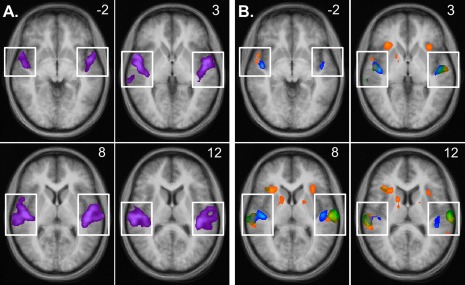
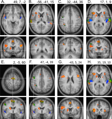
Neuroimaging studies investigating the voluntary (top-down) control of attention largely agree that this process recruits several frontal and parietal brain regions. Since most studies used attention tasks requiring several higher-order cognitive functions (e.g. working memory, semantic processing, temporal integration, spatial orienting) as well as different attentional mechanisms (attention shifting, distractor filtering), it is unclear what exactly the observed frontoparietal activations reflect. The present functional magnetic resonance imaging study investigated, within the same participants, signal changes in (1) a "Simple Attention" task in which participants attended to a single melody, (2) a "Selective Attention" task in which they simultaneously ignored another melody, and (3) a "Beep Monitoring" task in which participants listened in silence for a faint beep. Compared to resting conditions with identical stimulation, all tasks produced robust activation increases in auditory cortex, cross-modal inhibition in visual and somatosensory cortex, and decreases in the default mode network, indicating that participants were indeed focusing their attention on the auditory domain. However, signal increases in frontal and parietal brain areas were only observed for tasks 1 and 2, but completely absent for task 3. These results lead to the following conclusions: under most conditions, frontoparietal activations are crucial for attention since they subserve higher-order cognitive functions inherently related to attention. However, under circumstances that minimize other demands, nonspatial auditory attention in the absence of stimulation can be maintained without concurrent frontal or parietal activations.
Keywords: attention; auditory perception; baseline shift; default mode network; fMRI.
Copyright © 2013 Wiley Periodicals, Inc.
Figures


Similar articles
-
Brain activity during auditory and visual phonological, spatial and simple discrimination tasks.Brain Res. 2013 Feb 16;1496:55-69. doi: 10.1016/j.brainres.2012.12.013. Epub 2012 Dec 19. Brain Res. 2013. PMID: 23261663
-
Spatial attention evokes similar activation patterns for visual and auditory stimuli.J Cogn Neurosci. 2010 Feb;22(2):347-61. doi: 10.1162/jocn.2009.21241. J Cogn Neurosci. 2010. PMID: 19400684 Free PMC article.
-
Concurrent TMS-fMRI Reveals Interactions between Dorsal and Ventral Attentional Systems.J Neurosci. 2015 Aug 12;35(32):11445-57. doi: 10.1523/JNEUROSCI.0939-15.2015. J Neurosci. 2015. PMID: 26269649 Free PMC article.
-
Maps of space in human frontoparietal cortex.J Physiol Paris. 2013 Dec;107(6):510-6. doi: 10.1016/j.jphysparis.2013.04.002. Epub 2013 Apr 18. J Physiol Paris. 2013. PMID: 23603831 Free PMC article. Review.
-
New approaches to the study of human brain networks underlying spatial attention and related processes.Exp Brain Res. 2010 Oct;206(2):153-62. doi: 10.1007/s00221-010-2205-7. Epub 2010 Mar 31. Exp Brain Res. 2010. PMID: 20354681 Free PMC article. Review.
Cited by
-
Melodic Contour Identification Reflects the Cognitive Threshold of Aging.Front Aging Neurosci. 2016 Jun 13;8:134. doi: 10.3389/fnagi.2016.00134. eCollection 2016. Front Aging Neurosci. 2016. PMID: 27378907 Free PMC article.
-
Auditory-limbic interactions in chronic tinnitus: Challenges for neuroimaging research.Hear Res. 2016 Apr;334:49-57. doi: 10.1016/j.heares.2015.08.005. Epub 2015 Aug 20. Hear Res. 2016. PMID: 26299843 Free PMC article. Review.
-
Eye Movements during Auditory Attention Predict Individual Differences in Dorsal Attention Network Activity.Front Hum Neurosci. 2016 May 9;10:164. doi: 10.3389/fnhum.2016.00164. eCollection 2016. Front Hum Neurosci. 2016. PMID: 27242465 Free PMC article.
-
Manipulation of low-level features modulates grouping strength of auditory objects.Psychol Res. 2021 Sep;85(6):2256-2270. doi: 10.1007/s00426-020-01391-4. Epub 2020 Jul 20. Psychol Res. 2021. PMID: 32691138
-
Attentional Modulation of Hierarchical Speech Representations in a Multitalker Environment.Cereb Cortex. 2021 Oct 1;31(11):4986-5005. doi: 10.1093/cercor/bhab136. Cereb Cortex. 2021. PMID: 34115102 Free PMC article.
References
-
- Alain C, He Y, Grady C (2008): The contribution of the interior parietal lobe to auditory spatial working memory. J Cogn Neurosci 20:285–295. - PubMed
-
- Alho K, Medvedev SV, Pakhomov SV, Roudas MS, Tervaniemi M, Reinkainen K, Zeffiro T, Näätänen R (1999): Selective tuning of the left and right auditory cortices during spatially directed attention. Cogn Brain Res 7:335–341. - PubMed
-
- Alho K, Teder W, Lavikainen J, Näätänen R (1994): Strongly focused attention and auditory event‐related potentials. Biol Psychol 38:73–90. - PubMed
-
- Aron RA, Robbins TW, Poldrack RA (2004): Inhibition and the right inferior frontal cortex. Trends Cogn Sci 8:170–177. - PubMed
Publication types
MeSH terms
Substances
Grants and funding
LinkOut - more resources
Full Text Sources
Other Literature Sources
