. 2013 Nov;242(11):1320-31.
doi: 10.1002/dvdy.24021. Epub 2013 Oct 7.

A staging system for the regeneration of a polyp from the aboral physa of the anthozoan Cnidarian Nematostella vectensis

Affiliations

A staging system for the regeneration of a polyp from the aboral physa of the anthozoan Cnidarian Nematostella vectensis

Patricia E Bossert et al. Dev Dyn. 2013 Nov.

Abstract

Background: As the sea anemone Nematostella vectensis emerges as a model for studying regeneration, new tools will be needed to assess its regenerative processes and describe perturbations resulting from experimental investigation. Chief among these is the need for a universal set of staging criteria to establish morphological landmarks that will provide a common format for discussion among investigators.

Results: We have established morphological criteria to describe stages for rapidly assessing regeneration of the aboral end (physa) of Nematostella. Using this staging system, we observed rates of regeneration that are temperature independent during wound healing and temperature dependent afterward. Treatment with 25 μM lipoic acid delays the progression through wound healing without significantly affecting the subsequent rate of regeneration. Also, while an 11-day starvation before amputation causes only a minimal delay in regeneration, this delay is exacerbated by lipoic acid treatment.

Conclusions: A system for staging the progression of regeneration in amputated Nematostella physa based on easily discernible morphological features provides a standard for the field. This system has allowed us to identify both temperature-dependent and -independent phases of regeneration, as well as a nutritional requirement for normal regenerative progression that is exacerbated by lipoic acid.

Keywords: Anthozoa; Cnidarian; Nematostella; lipoic acid; regeneration; staging system.


Figures

Figure 1
Figure 1. Nematostella Anatomy
(A) Photograph showing a Nematostella vectensis polyp. Dotted red line indicates amputation level. Scale bar is 0.5 mm. (B) Cartoon illustrating several anatomical structures of the polyp. Capitulum, scapus, and physa are distinguished, in addition to axial location, by ectodermal thickness and organization. Wavy light gray lines correspond to tentacles, dark gray spots are nematocysts, the orange trapezoid represents the pharynx, thick black lines are the mesenterial insertions, the dark purple bars are the mesenteries. (C) Cartoon cross section of the dotted oval in B, illustrating the radial projection of the eight mesenteries. (D) High magnification cartoon of the area in the dotted rectangle in (B) illustrating pleated mesentery as seen from an oblique angle. (E) High magnification cartoon of a mesentery viewed in cross section. Muscle (diagonal lines) tissue is located closest to the body wall, gonads (white circles) are positioned medially, and the digestive region, including the trefoil-shaped mesenterial filament, is located at the free edge of the mesentery, within the coelenteron (Stephenson, 1928a). (apt) aperture, (cap) capitulum, (ent) enterostome, (mf) mesenterial filament, (mi) mesenterial insertion, (ph) physa, (sc) scapus.
Figure 2
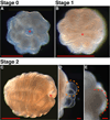
Figure 2. NRSS Stages 0 through 2
(A) Stage 0, Open Wound. The wound site may be visibly expanding and contracting, and is seen here in a contracted position. (B) Stage 1, Closed Wound. The wound has closed, although the mesenterial insertions may not meet in an organized fashion at the new oral pole. (C) Stage 2, Radial Arches. The puffed arches, appearing constrained at the mesenterial insertions, give the oral pole the appearance of a stalk-less pumpkin. (D) High magnification view of tissue blebs (dotted orange line). The bleb is nearly spherical in shape and is a single cell layer thick. (E) High magnification view of tentacle buds (dotted red line). Tenacle buds are wider at the base than tip, may appear mound-shaped or pointed, and are two cell layers thick. (A) and (B) are oriented with the oral pole facing out from the page. (C–E) are oriented with the oral pole to the right. Asterisks indicate site of amputation/oral pole. Scale bars are 0.5mm (A–C) and 0.1mm (D,E).
Figure 3
Figure 3. NRSS Stages 3 through 5
(A–E) Stage 3, Tentacles. (A) Tentacle rudiments, longer than wide, are visible at the oral pole. Both pharynx (orange arrowhead) and regenegesta (green arrowhead) may be visible at this stage. (B) High magnification view of the box in (A) illustrating the tentacle bud. (C) The ratio of the oral-aboral axis to the directive axis is variable at this stage, as the animals begin to inflate or contract along their axes. (D) Early mesentery primordia may be visible in Stage 3, but recede into the mesenterial insertion at a distance of less than twice the height from enterostome to mesenterial insertion. Purple arrowhead indicates mesentery. Green and orange arrowheads are as in (A). (E) High magnification view of the box in (D) illustrating the recession of the mesenterial filament into the mesenterial insertion. (F–J) Stage 4, Linear Mesentery. (F) Recession of the mesenterial filament into the mesenterial insertion extends beyond twice the distance from the enterstome to the mesenterial insertion. Arrowheads are as in (D). (G) High magnification view of the box in (F) showing the increased length of the mesentery compared to (E). (H) Oral-aboral axis length, scapus opacity, and tentacle number and length are variable. (I) High magnification view of the box in (H) illustrating early pleating of mesenteries (green line) in the same animal with unpleated mesenteries (yellow line). (J) Aboral view of a Stage 4 animal with four pleated (green arrowheads) and four unpleated (yellow arrowheads) mesenteries. (K–M) Stage 5, Pleated Mesentery. (K) Aboral view of a Stage 5 animal with 8 pleated mesenteries (green arrowheads). Note that asymmetric inflation diminishes apparent pleating in one mesentery (black arrowhead). (L) The majority of the mesenteries demonstrate visible pleating. (M) High magnification view of the box in (L) illustrating the increased extent of pleating (green line). Scale bars are 0.5mm.
Figure 4
Figure 4. Cartoon Illustrating Progression of NRSS Stages
(A) Stage 0, open wound. (B) Stage 0 - double-headed red arrows indicate the dynamic expansion and contraction of the wound edge (dotted circle). (C) Stage 1, wound closure (red arrows) at new oral pole (green arrowhead). (D) Stage 1 with additional morphologies: the oral pole appears to become recessed relative to the apex of the inflating arches. (E) Stage 2, arching of oral tissue results in pumpkinhead appearance (green arrowhead). (F) Stage 2 with additional morphologies: physa may begin to elongate and narrow (red arrows). Tentacle buds (red) and blebs (light blue) may appear at the oral ring. (G) Stage 3, tentacles (green arrowhead) appear longer than wide. (H) Stage 3 with additional morphologies: darkening of tissue (orange mass) corresponding to regenerating pharynx may be visible (green arrowhead). (I and I') Stage 4, linear and unpleated mesenteries appear at least twice as long as they are tall at the enterostome. Mesenteries of sufficient (check) and insufficient ('X') length are diagrammed. (J and J') Stage 4 with additional morphologies: mesenterial filaments acquire a pleated appearance in four or less mesenteries. Tentacles increase in number and length. (K) Stage 5, pleated mesenteries now number more than four. (K') Aboral view of Stage 5 mesenteries. All animals oriented with oral pole to the right, except in K'.
Figure 5
Figure 5. Inter-Rater Reliability of the NRSS
Graph comparing Average Stage Regeneration of four sets of five amputated physa at 18 °C, as scored by MPD and PEB. Investigators were scoring the same animals independently without communication.
Figure 6
Figure 6. Effects of Temperature on Regeneration
(A) Initial wound healing stages of regeneration appear temperature independent, while the rate of regeneration through later stages varies with culture temperature. Error bars correspond to standard error of the mean, n=4. (B) The number of regenegesta observed throughout regeneration varied with temperature. Note that the y-axis corresponds to the number of regenegesta observed each day and is not cumulative.
Figure 7
Figure 7. Effects of Lipoic Acid and Starvation on Regeneration
Physa treated with lipoic acid, prior starvation, or both, exhibited delays in regeneration compared to untreated control physa. Lipoic acid treatment, regardless of prior nutritional state, results in a lag in progression from Stage 1 to Stage 2. Prior starvation alone causes a 6–18 hour delay in stage progression beginning at Stage 2, while combination of both lipoic acid and prior starvation exacerbates this lag. Error bars correspond to standard error of the mean, n=4.

References

    1. Agata K, Saito Y, Nakajima E. Unifying principles of regeneration I: Epimorphosis versus morphallaxis. Develop. Growth Differ. 2007;49:73–78. - PubMed
    1. Agata K, Tanaka T, Kobayashi C, Kato K, Saitoh Y. Intercalary Regeneration in Planarians. Developmental Dynamics. 2003;226:308–316. - PubMed
    1. Ball EE, Hayward DC, Saint R, Miller DJ. A simple plan - cnidarians and the origins of developmental mechanisms. Nature Reviews Genetics. 2004;5:567–577. - PubMed
    1. Bossert P, Galliot B. How to use Hydra as a model system to teach biology in the classroom. International Journal of Developmental Biology. 2012;56:637–652. - PubMed
    1. Brøndsted A, Brøndsted HV. Influence of Temperature on Rate of Regeneration in the Time-graded Regeneration Field in Planarians. Journal of Embryology and Experimental Morphology. 1961;9:159–166.

Publication types

LinkOut - more resources