. 2013 Nov;27(11):4444-54.
doi: 10.1096/fj.12-224907. Epub 2013 Aug 2.

Profibrotic TGFβ responses require the cooperative action of PDGF and ErbB receptor tyrosine kinases

Affiliations

Profibrotic TGFβ responses require the cooperative action of PDGF and ErbB receptor tyrosine kinases

Mahefatiana Andrianifahanana et al. FASEB J. 2013 Nov.

Abstract

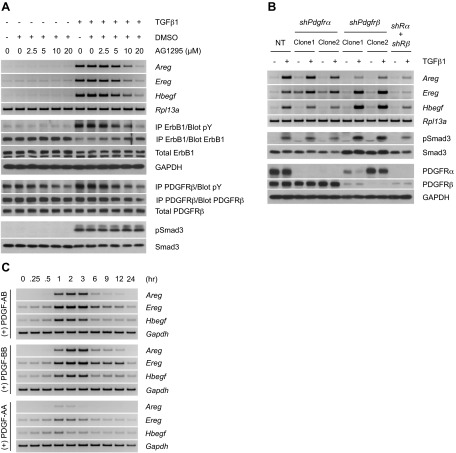
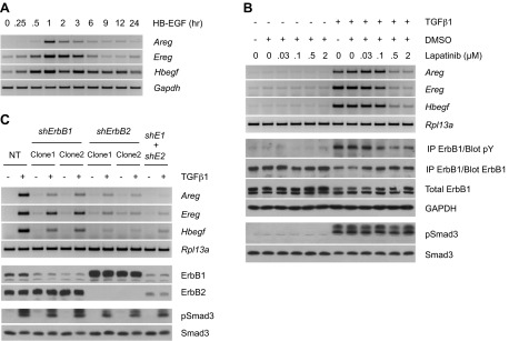
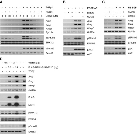
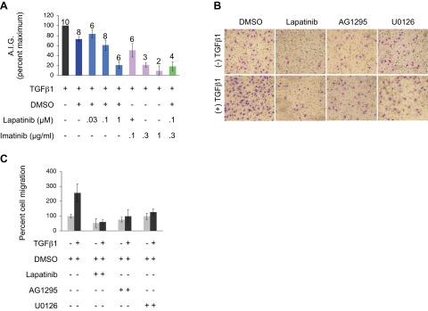
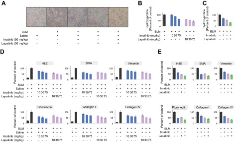
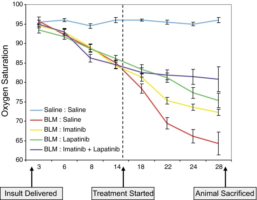
Transforming growth factor β (TGFβ) has significant profibrotic activity both in vitro and in vivo. This reflects its capacity to stimulate fibrogenic mediators and induce the expression of other profibrotic cytokines such as platelet-derived growth factor (PDGF) and epidermal growth factor (EGF/ErbB) ligands. Here we address both the mechanisms by which TGFβ induced ErbB ligands and the physiological significance of inhibiting multiple TGFβ-regulated processes. The data document that ErbB ligand induction requires PDGF receptor (PDGFR) mediation and engages a positive autocrine/paracrine feedback loop via ErbB receptors. Whereas PDGFRs are essential for TGFβ-stimulated ErbB ligand up-regulation, TGFβ-specific signals are also required for ErbB receptor activation. Subsequent profibrotic responses are shown to involve the cooperative action of PDGF and ErbB signaling. Moreover, using a murine treatment model of bleomycin-induced pulmonary fibrosis we found that inhibition of TGFβ/PDGF and ErbB pathways with imatinib plus lapatinib, respectively, not only prevented myofibroblast gene expression to a greater extent than either drug alone, but also essentially stabilized gas exchange (oxygen saturation) as an overall measure of lung function. These observations provide important mechanistic insights into profibrotic TGFβ signaling and indicate that targeting multiple cytokines represents a possible strategy to ameliorate organ fibrosis dependent on TGFβ.

Keywords: EGF; pulmonary fibrosis.


Figures

Figure 1.
Figure 1.
TGFβ induces ErbB signaling via autocrine activation of the PDGFR pathway. A) RT-PCR analyses of ErbB ligands (12 h post-treatment) and Western blot analysis of ErbB1 phosphorylation (15 h) following cell stimulation with TGFβ1 in the presence of the PDGFR inhibitor AG1295. The profile of PDGFR phosphorylation (12 h) confirms the efficacy of AG1295, whereas that of Smad3 (3 h) proves its specificity. B) RT-PCR analyses of ErbB ligands (12 h post-treatment) upon TGFβ1 stimulation of cells transduced with shRNA targeting PDGFRα, PDGFRβ, or both (shRα+shRβ). NT denotes nontargeting control. Western blot analyses show consistent Smad3 phosphorylation and efficient knockdown of PDGFRα and PDGFRβ (3 h). The data presented further corroborate the role of PDGFR in mediating up-regulation of ErbB ligands by TGFβ. C) Time course of ErbB ligand induction by PDGF in AKR-2B cells. RT-PCR analyses of ErbB ligands show a fairly rapid up-regulation of ErbB ligands by PDGF isoforms, with isoforms containing the B chain being the most potent.
Figure 2.
Figure 2.
TGFβ stimulates ErbB ligand expression via ErbB autoinduction. A) RT-PCR analyses of ErbB ligand expression in HB-EGF-treated AKR-2B cells. Activation of ErbB induces expression of its own ligands. B) RT-PCR analyses of ErbB ligands and Western blot analysis of ErbB1 phosphorylation (15 h post-treatment) following cell stimulation with TGFβ1 in the presence of the ErbB1/ErbB2 inhibitor, lapatinib. The lack of effect on Smad3 phosphorylation (3 h) confirms the specificity of lapatinib. Stimulation of ErbB ligand expression by TGFβ requires ErbB autoinduction. C) RT-PCR analyses of ErbB ligands (12 h post-treatment) in response to TGFβ1 stimulation of cells stably expressing shRNA targeting ErbB1, ErbB2, or both (shE1+shE2). Western blot analyses (1 h) show efficient knockdown of ErbB1 and ErbB2 and uniform activation of Smad3. Results further support the role of ErbB autoinduction in ErbB ligand up-regulation by TGFβ.
Figure 3.
Figure 3.
Up-regulation of ErbB ligands by TGFβ requires MEK activation. A) RT-PCR analyses of ErbB ligands (12 h post-treatment) on TGFβ1 stimulation of cells in the presence of the MEK inhibitor, U0126. Inhibition of TGFβ1-stimulated ERK1/2 phosphorylation (9 h) demonstrates the efficacy of U0126, and the lack of effect on Smad3 phosphorylation (3 h) verifies its specificity. Induction of ErbB ligands by TGFβ is mediated by MEK. B, C) RT-PCR analyses of ErbB ligands following stimulation of cells with PDGF-AB (2 h post-treatment) or HB-EGF (1 h post-treatment) in the presence of U0126. Western blot analyses of ERK1/2 and AKT phosphorylation (15 min) confirm drug efficacy and specificity, respectively. PDGFR- and ErbB-driven ErbB ligand up-regulation is MEK-dependent. D) RT-PCR analyses of ErbB ligands in cells transfected with control vector or FLAG-tagged constitutively active MEK1 (FLAG-MEK1-S218-222D) for 48 h. TGFβ1-stimulated cells (12 h) were used as a positive control for ErbB ligand induction. Phosphorylation of the downstream targets ERK1/2 indicates MEK activation. The results support the notion that activation of MEK is sufficient for ErbB ligand induction.
Figure 4.
Figure 4.
TGFβ stimulates the ErbB pathway through integration of autocrine PDGFR signals and additional TGFβ-specific input. A) Western blot analysis of ErbB1 phosphorylation following stimulation of cells with TGFβ1 or PDGF isoforms. Whereas both TGFβ and PDGF are able to induce ErbB ligand expression, only TGFβ can trigger ErbB phosphorylation. B) Western blot analyses showing phosphorylation of ERK1/2 by both PDGF isoforms (15 min). C) ELISA assay of conditioned media from cells stimulated with TGFβ1 or PDGF isoforms. Equivalent aliquots of cell lysates from 6 replicates used in ELISA were analyzed by Western blotting for β-tubulin expression. Data shown are from one representative experiment, repeated twice. Error bars = sd. Only TGFβ, but not PDGF, is capable of inducing EREG shedding. Results presented in A–C demonstrate that PDGFR activation is necessary and sufficient for TGFβ-induced ErbB ligand up-regulation, but not ErbB phosphorylation. D) Proposed model for profibrotic TGFβ signaling. Engagement of the TGFβ receptor (TGFβR) complex activates the canonical Smad pathway, leading to up-regulation of PDGFR ligands. Subsequent PDGFR phosphorylation promotes activation of the MEK pathway and stimulates ErbB ligand up-regulation. Additional TGFβR signals facilitate shedding of ErbB ligands and activation of cognate receptors, which further generate a positive feedback loop to induce its own ligands via MEK. Question marks denote potential mechanisms whereby TGFβ may independently regulate sheddase activity or, alternatively, control translation of ErbB ligand message. The PDGFR and ErbB pathways, in concert with the noncanonical TGFβ-activated signaling modules PAK2/cAbl and PI3K/AKT/mTOR (19–23, 25, 26, 29), all contribute to the fibrogenic program directed by TGFβ.
Figure 5.
Figure 5.
TGFβ-induced anchorage-independent growth (AIG) and migration require PDGFR and ErbB mediation. A) Analyses of TGFβ1-stimulated AIG in the presence of lapatinib and/or imatinib. Treatments were done in triplicate; number of experiments included for each particular treatment is indicated. Error bars = sem. Significant inhibition of ErbB and PDGFR can be attained using higher concentrations of lapatinib and imatinib, respectively, whereas a combination of both drugs at suboptimal doses yields a synergistic effect. B) Analyses of TGFβ1-induced cell migration in the presence of pharmacological inhibitors of ErbB (lapatinib; 5 μM), PDGFR (AG1295; 20 μM), and MEK (U0126; 10 μM). Representative photomicrographs are shown. C) Quantitative analysis of B, where values are from 2 independent experiments, with 4 replicates/treatment. Error bars = sd. Results in B and C show that all 3 pathways are required for TGFβ-induced cell migration.
Figure 6.
Figure 6.
Imatinib and lapatinib cooperate to attenuate BLM-induced lung remodeling. A) Masson's Trichrome staining of paraffin-embedded lung sections from mice challenged with BLM (or saline) for 28 d and treated daily with imatinib and/or lapatinib beginning 15 d following initial BLM insult. Representative photomicrographs are shown (original view ×10). B, C) Quantitative analysis of hydroxyproline contents of lung sections generated from mice treated as in A. D, E) Pixellation scores (expressed as percentage of positive BLM control; ref. 30) quantifying H&E or immunohistochemical staining of lung sections prepared as in A. Data show expression or deposition of smooth muscle actin (SMA), vimentin, fibronectin, collagen I or collagen IV. H&E staining facilitates a direct visualization of collagen. Errors bars = sd. For C and E, both imatinib and lapatinib were used at 50 mg/kg/d. Results support a cooperative therapeutic role of imatinib and lapatinib in attenuating lung remodeling, as evidenced by reduced extracellular matrix production/deposition.
Figure 7.
Figure 7.
Dual therapy with imatinib and lapatinib stabilizes lung gas exchange in BLM-challenged mice. Time-dependent fluctuation of oxygen saturation (SpO2) levels (determined on room air) in mice challenged with BLM (or saline) for 28 d and treated with imatinib and/or lapatinib (both at 50 mg/kg/d) beginning 15 d after initial BLM insult. Errors bars = sem (n=6). While administration of individual drugs resulted in partial recovery, a dual treatment yielded a cooperative effect on lung physiology, as indicated by the respective increases in dissolved oxygen levels.

References

    1. Steele M. P., Schwartz D. A. (2012) Molecular mechanisms in progressive idiopathic pulmonary fibrosis. Annu. Rev. Med. 64, 265–276 - PubMed
    1. Todd N. W., Luzina I. G., Atamas S. P. (2012) Molecular and cellular mechanisms of pulmonary fibrosis. Fibrogenesis Tissue Repair 5, 11. - PMC - PubMed
    1. Wynn T. A. (2007) Common and unique mechanisms regulate fibrosis in various fibroproliferative diseases. J. Clin. Invest. 117, 524–529 - PMC - PubMed
    1. Wynn T. A., Ramalingam T. R. (2012) Mechanisms of fibrosis: therapeutic translation for fibrotic disease. Nat. Med. 18, 1028–1040 - PMC - PubMed
    1. Sivakumar P., Ntolios P., Jenkins G., Laurent G. (2012) Into the matrix: targeting fibroblasts in pulmonary fibrosis. Curr. Opin. Pulm. Med. 18, 462–469 - PubMed

Publication types

MeSH terms