. 2013 Nov;23(11):1789-96.
doi: 10.1101/gr.161265.113. Epub 2013 Aug 2.

RNA-DNA differences in human mitochondria restore ancestral form of 16S ribosomal RNA

Affiliations

RNA-DNA differences in human mitochondria restore ancestral form of 16S ribosomal RNA

Dan Bar-Yaacov et al. Genome Res. 2013 Nov.

Abstract

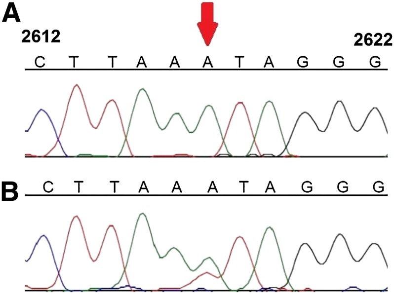
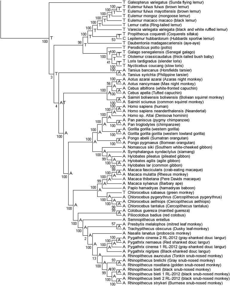
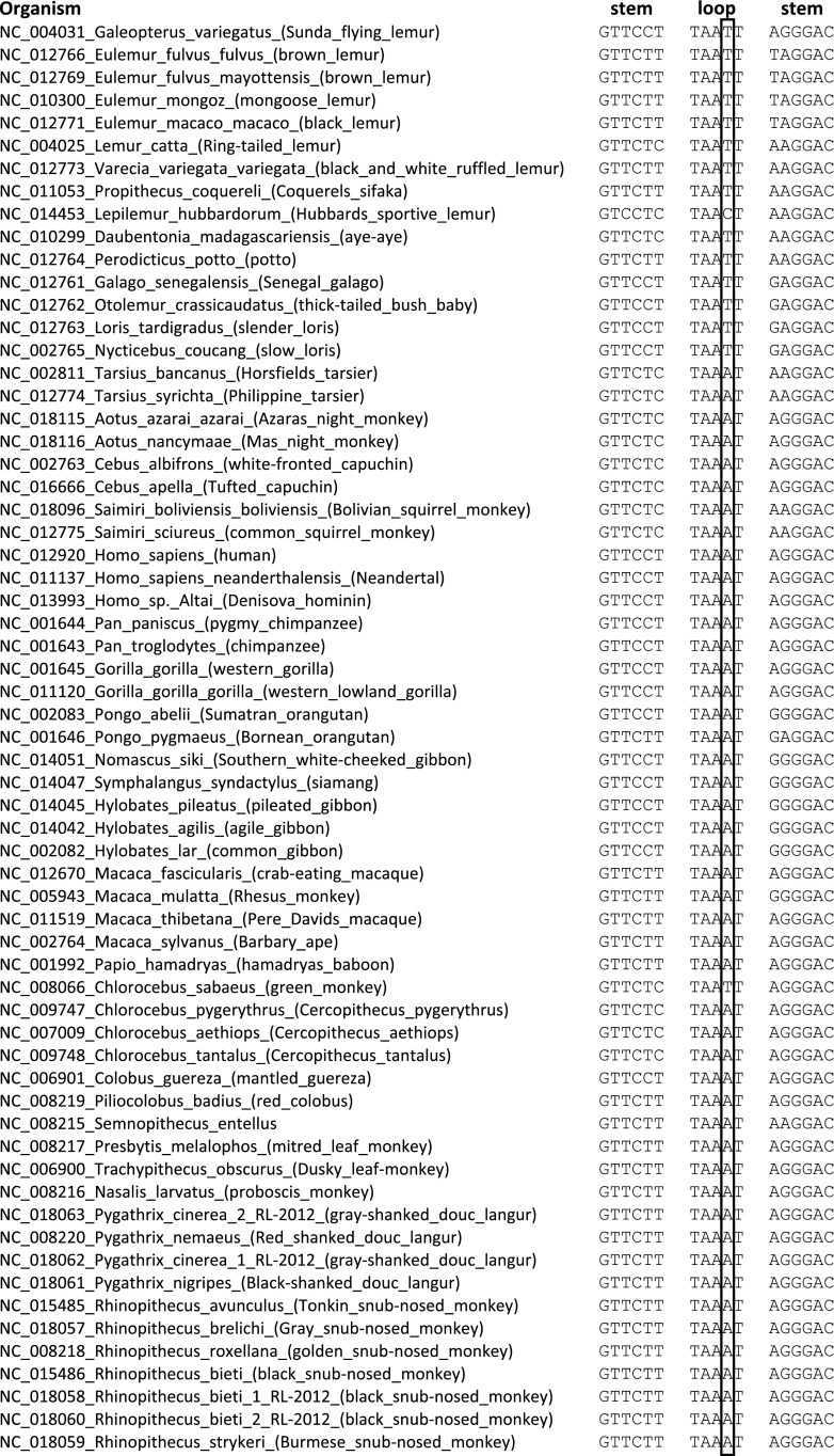
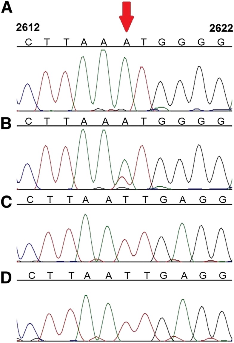
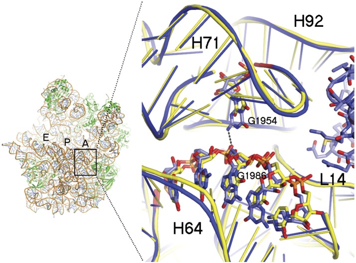
RNA transcripts are generally identical to the underlying DNA sequences. Nevertheless, RNA-DNA differences (RDDs) were found in the nuclear human genome and in plants and animals but not in human mitochondria. Here, by deep sequencing of human mitochondrial DNA (mtDNA) and RNA, we identified three RDD sites at mtDNA positions 295 (C-to-U), 13710 (A-to-U, A-to-G), and 2617 (A-to-U, A-to-G). Position 2617, within the 16S rRNA, harbored the most prevalent RDDs (>30% A-to-U and ∼15% A-to-G of the reads in all tested samples). The 2617 RDDs appeared already at the precursor polycistrone mitochondrial transcript. By using traditional Sanger sequencing, we identified the A-to-U RDD in six different cell lines and representative primates (Gorilla gorilla, Pongo pigmaeus, and Macaca mulatta), suggesting conservation of the mechanism generating such RDD. Phylogenetic analysis of more than 1700 vertebrate mtDNA sequences supported a thymine as the primate ancestral allele at position 2617, suggesting that the 2617 RDD recapitulates the ancestral 16S rRNA. Modeling U or G (the RDDs) at position 2617 stabilized the large ribosomal subunit structure in contrast to destabilization by an A (the pre-RDDs). Hence, these mitochondrial RDDs are likely functional.


Figures

Figure 1.
Figure 1.
Validation of the RDD at position 2617 by Sanger sequencing. The numbers from each side of the sequences correspond to mtDNA positions. Red arrow points at position 2617. Presented is a representative sample: cultured B cells from GM14468.
Figure 2.
Figure 2.
Phylogenetic analysis of mitochondrial DNA sequences of 62 primates and one Dermoptera (Galeopterus variegatus). The primate portion of the phylogenetic analysis of mtDNA eutherian sequences from 334 organisms; the full vertebrate tree is shown in Supplemental Figure 6. Numbers on the branches are scores from 1000 bootstrap replicates. Ancestral state of position 2617 is indicated for each branch.
Figure 3.
Figure 3.
Multiple sequence alignment of mitochondrial DNA sequences of 62 primates and one Dermoptera (G. variegatus). Shown are primate orthologs of human mtDNA positions 2608–2624 (framed is the nucleotide at position 2617). Stem, Loop, Stem columns indicate the mtDNA sequence corresponding to the stem-and-loop structure of 16S rRNA around position 2617. The full vertebrate sequence alignment is in Supplemental Figure 5.
Figure 4.
Figure 4.
Sanger sequence analysis of position 2617 from DNA and RNA samples of mtDNA from various primates. Pongo pigmaeus (A) DNA and (B) RNA; Nycticebus coucang (C) DNA and (D) RNA. Red arrow indicates nucleotide position corresponding to position 2617 in human mtDNA. Numbers on top of each base indicate positions according to the rCRS.
Figure 5.
Figure 5.
Structure of the ribosome section corresponding to region orthologous to that of position 2617. (Left) The large ribosomal subunit from a bacterium, Deinococcus radiodurans, represented as ribbon. The A-, P-, and E-binding sites of tRNA on the ribosomal large subunit are shown. (Right) Sticks-and-ribbon representation of H71 and H64 interaction in the bacterial (blue) or human (yellow) orthologous ribosomes. The hydrogen bond that is disrupted by an adenine in position 2617 is represented as a dashed line. Numbers represent positions of E. coli ribosomal RNA.

References

    1. Anger AM, Armache JP, Berninghausen O, Habeck M, Subklewe M, Wilson DN, Beckmann R 2013. Structures of the human and Drosophila 80S ribosome. Nature 497: 80–85 - PubMed
    1. Avital G, Buchshtav M, Zhidkov I, Tuval Feder J, Dadon S, Rubin E, Glass D, Spector TD, Mishmar D 2012. Mitochondrial DNA heteroplasmy in diabetes and normal adults: Role of acquired and inherited mutational patterns in twins. Hum Mol Genet 21: 4214–4224 - PMC - PubMed
    1. Ben-Shem A, Garreau de Loubresse N, Melnikov S, Jenner L, Yusupova G, Yusupov M 2011. The structure of the eukaryotic ribosome at 3.0 A resolution. Science 334: 1524–1529 - PubMed
    1. Bentley DR, Balasubramanian S, Swerdlow HP, Smith GP, Milton J, Brown CG, Hall KP, Evers DJ, Barnes CL, Bignell HR, et al. 2008. Accurate whole human genome sequencing using reversible terminator chemistry. Nature 456: 53–59 - PMC - PubMed
    1. Bestwick ML, Shadel GS 2013. Accessorizing the human mitochondrial transcription machinery. Trends Biochem Sci 38: 283–291 - PMC - PubMed

Publication types