Review
. 2013 Nov;64(15):4663-80.
doi: 10.1093/jxb/ert204. Epub 2013 Aug 3.

A force of nature: molecular mechanisms of mechanoperception in plants

Affiliations
Review

A force of nature: molecular mechanisms of mechanoperception in plants

Gabriele B Monshausen et al. J Exp Bot. 2013 Nov.

Abstract

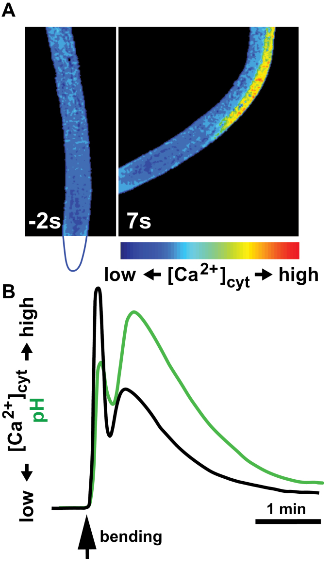
The ability to sense and respond to a wide variety of mechanical stimuli-gravity, touch, osmotic pressure, or the resistance of the cell wall-is a critical feature of every plant cell, whether or not it is specialized for mechanotransduction. Mechanoperceptive events are an essential part of plant life, required for normal growth and development at the cell, tissue, and whole-plant level and for the proper response to an array of biotic and abiotic stresses. One current challenge for plant mechanobiologists is to link these physiological responses to specific mechanoreceptors and signal transduction pathways. Here, we describe recent progress in the identification and characterization of two classes of putative mechanoreceptors, ion channels and receptor-like kinases. We also discuss how the secondary messenger Ca(2+) operates at the centre of many of these mechanical signal transduction pathways.

Keywords: Calcium; cell-wall integrity; mechanoperception; mechanosensitive ion channels; receptor-like kinases; thigmomorphogenesis..


Figures

Fig. 1.
Fig. 1.
External and internal mechanical forces cause deformation (strain) of plant cells. When a plant organ is bent (top), cells on the convex side are stretched (experience positive strain) while cells on the opposite, concave side are compressed (negative strain). During rapid turgor-driven cell expansion (bottom), local positive strain rates as high as 50–70% h–1 have been measured in the elongation zone of maize and Arabidopsis roots (Ishikawa and Evans, 1993; G. Monshausen and N. Miller, unpublished data).
Fig. 2.
Fig. 2.
Model of mechanosensing and signal transduction. Mechanosensor proteins are activated when they undergo a conformational change in response to a mechanical force. Ion channels such as MSLs and putative stretch-activated Ca2+-permeable channels (SACC) such as MCA or Piezo are gated by changes in membrane tension. Other mechanosensory proteins may be linked to intra- and/or extracellular tethers such as the cytoskeleton or glycosylated proteins and polysaccharides of the cell wall; mechanical forces acting on sensors through these linkages could cause conformational changes by breaking or stabilizing intra- and intermolecular bonds (e.g. protein unfolding, catch bonds; Vogel and Sheetz, 2006). Receptor-like kinases with (putative) carbohydrate-binding domains are found among the CrRLK1L, WAKs, S-domain, and lectin-like RLK subfamilies (Gish and Clark, 2011) and may transmit information about deformation of the cell wall to the cell interior via kinase-dependent phosphorylation of target proteins. Downstream targets could include transcription factors (TF-P) to regulate the expression of mechanoresponsive genes or Ca2+-permeable channels (CC) that, in conjunction with SACC, would shape the specific signature of mechanically triggered Ca2+ signals. [Ca2+]cyt changes are typically interpreted by the Ca2+ sensors calmodulin (CaM) and calmodulin-like proteins, Ca2+-dependent protein kinases (CDPKs), and calcineurin B-like proteins (CBLs) (Hashimoto and Kudla, 2011) or directly by target proteins harbouring Ca2+-binding motifs. Ca2+ signalling regulates the expression of (some) mechanoresponsive genes and may be linked to the biosynthesis of jasmonic acid, a key reglulator of plant thigmomorphogenesis. Ca2+ signalling also activates plasma membrane transport processes (e.g. NADPH-oxidase mediated reactive oxygen species production or H+/OH transport to alter apoplastic and cytosolic pH) that could rapidly alter cell-wall extensibility. Mechanical stress may also directly disrupt cell-wall pectate structure and weaken Ca2+–pectate cross-bridges to promote cell-wall remodelling (Boyer, 2009).
Fig. 3.
Fig. 3.
Simplified two-state models for the gating of MS ion channels. In the intrinsic model (A), the open state (that which conducts ions) of an MS channel, shaded in grey, is favoured by increased membrane tension, which leads to membrane thinning and/or to changes in the force exerted on the protein–lipid interface. Alternatively (B), the open state is favoured by the opening of a ‘trapdoor’ domain that is tethered to an elastic component of the cytoskeleton or cell wall (indicated by a black bar).
Fig. 4.
Fig. 4.
Single-channel patch-clamp analysis of MS channels expressed in Xenopus oocytes. A thin glass pipette is used to puncture a Xenopus oocyte, indicated by the dashed box in (A), capturing a patch of membrane in the tip, as shown in (B). Negative pressure (suction) introduced through the pipette deforms the patch of membrane, increasing membrane tension and gating intrinsically MS ion channels (C). A step-wise increase in current can be observed as individual channels present in the patch pipette open upon application of suction (D) (E. Haswell and G. Maksaev, unpublished data).
Fig. 5.
Fig. 5.
Ion signalling in roots in response to mechanical bending. (A) Arabidopsis root expressing the FRET-based Ca2+ biosensor yellow cameleon 3.6 (Monshausen et al., 2009) is bent to the side with the help of a glass capillary. The position of the root tip (not in the field of view) is outlined in blue below the left panel. Roots exhibit low resting [Ca2+]cyt prior to bending (left) and a rapid increase in [Ca2+]cyt after bending on the stretched (convex) side but not the compressed (concave) side of the roots (right). (B) Kinetics of mechanically triggered [Ca2+]cyt changes in root epidermal cells are echoed by the kinetics of changes in extracellular pH monitored using the fluorescent pH sensor fluorescein conjugated to dextran (based on Monshausen et al., 2009).

References

    1. Adamec L. 2012. Firing and resetting characteristics of carnivorous Utricularia reflexa traps: physiological or only physical regulation of trap triggering? Phyton-Annales Rei Botanicae 52, 281–290
    1. Alexandre J, Lassalles JP. 1991. Hydrostatic and osmotic pressure activated channel in plant vacuole. Biophysical Journal 60, 1326–1336 - PMC - PubMed
    1. Anten NPR, Alcala-Herrera R, Schieving F, Onoda Y. 2010. Wind and mechanical stimuli differentially affect leaf traits in Plantago major . New Phytologist 188, 554–564 - PubMed
    1. Arnadottir J, Chalfie M. 2010. Eukaryotic mechanosensitive channels. Annual Review of Biophysics 39, 111–137 - PubMed
    1. Balleza D, Gomez-Lagunas F. 2009. Conserved motifs in mechanosensitive channels MscL and MscS. European Biophysical Journal 38, 1013–1027 - PubMed

Publication types