. 2013 Oct 1:250:403-424.
doi: 10.1016/j.jcp.2013.05.027.

Inverse Electrocardiographic Source Localization of Ischemia: An Optimization Framework and Finite Element Solution

Affiliations

Inverse Electrocardiographic Source Localization of Ischemia: An Optimization Framework and Finite Element Solution

Dafang Wang et al. J Comput Phys. .

Abstract

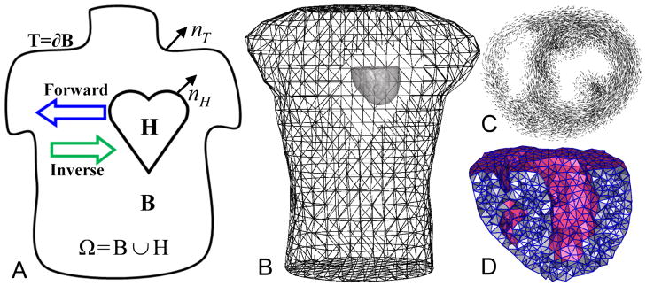
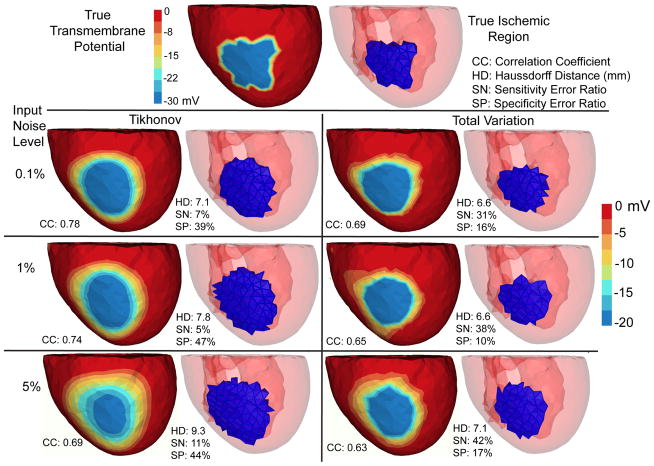
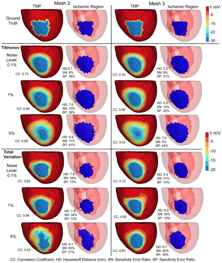
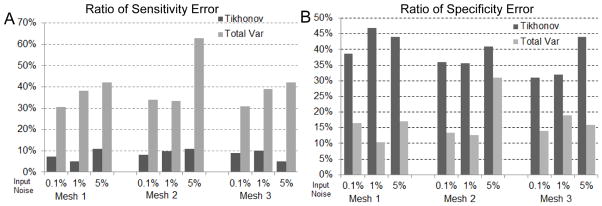
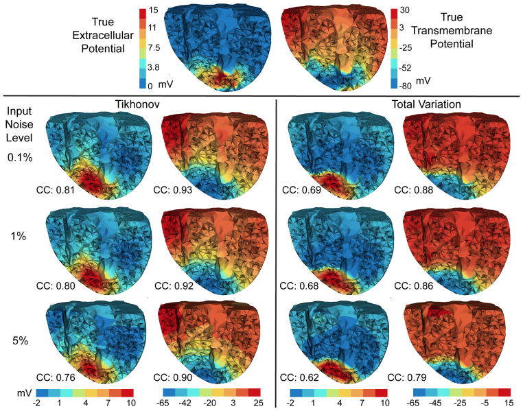
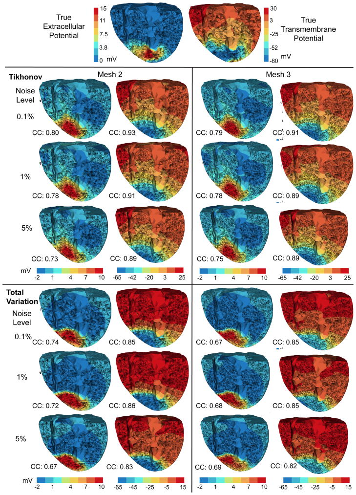
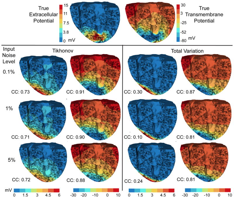
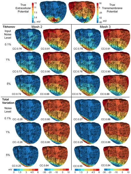
With the goal of non-invasively localizing cardiac ischemic disease using body-surface potential recordings, we attempted to reconstruct the transmembrane potential (TMP) throughout the myocardium with the bidomain heart model. The task is an inverse source problem governed by partial differential equations (PDE). Our main contribution is solving the inverse problem within a PDE-constrained optimization framework that enables various physically-based constraints in both equality and inequality forms. We formulated the optimality conditions rigorously in the continuum before deriving finite element discretization, thereby making the optimization independent of discretization choice. Such a formulation was derived for the L2-norm Tikhonov regularization and the total variation minimization. The subsequent numerical optimization was fulfilled by a primal-dual interior-point method tailored to our problem's specific structure. Our simulations used realistic, fiber-included heart models consisting of up to 18,000 nodes, much finer than any inverse models previously reported. With synthetic ischemia data we localized ischemic regions with roughly a 10% false-negative rate or a 20% false-positive rate under conditions up to 5% input noise. With ischemia data measured from animal experiments, we reconstructed TMPs with roughly 0.9 correlation with the ground truth. While precisely estimating the TMP in general cases remains an open problem, our study shows the feasibility of reconstructing TMP during the ST interval as a means of ischemia localization.

Keywords: Bidomain Model; Electrocardiography; Finite Element Method; Inverse Problem; Myocardial Ischemia; PDE Optimization; Total Variation.


Figures

Fig. 1
Fig. 1
Panel (A): the problem domain. (B): the heart/torso geometry. (C): fiber structure of a 1 cm-thick slice of the heart. (D): a cross-sectional view of the heart mesh.
Fig. 2
Fig. 2
Inverse solutions based on Mesh 1 with synthetic ischemia data, obtained by the Tikhonov and the total variation methods. Ischemic regions are indicated by the blue color. All the calculated TMPs are in the same color scale. This figure is to be considered with Fig 4. The input noise is i.i.d. Gaussian imposed on each node on the body surface. The noise level indicates the noise-to-signal ratio in the root-mean-square sense. Such noise setting holds for all experiments in this paper.
Fig. 3
Fig. 3
Inverse solutions based on Mesh 2 and Mesh 3, with synthetic ischemia data. Tikhonov and total variation solutions are presented. TMP stands for the transmembrane potential. Ischemic regions are denoted by the blue region. All the calculated TMPs are in the same color scale. This figure is to be compared with Fig 2 and Fig 4. The mesh profiles are given in Table 1.
Fig. 4
Fig. 4
Accuracy of ischemia localization with synthetic ischemia data, in terms of the ratios of sensitivity error (Panel A) and of specificity error (Panel B). This figure is to be compared with Fig 2 and Fig 3.
Fig. 5
Fig. 5
Inverse solutions based on Mesh 1 using the anisotropic inverse model and measured ischemia data. Reconstructed heart potentials (by Tikhonov and total variation methods) are visualized at the same cross section. Figures in each column are in the same color scale. CC denotes the correlation coefficient between each reconstructed potential and the ground truth (the top row).
Fig. 6
Fig. 6
Inverse solutions based on Mesh 2 and Mesh 3, using the anisotropic model and the measured ischemia data. Each column of reconstructed heart potentials are in the same color scale. CC: the correlation coefficient between the computed potential and the ground truth (the top row).
Fig. 7
Fig. 7
Inverse computation time of the Tikhonov (left) and the total variation regularization (right) based on the anisotropic heart model and measured ischemia data (shown by Fig 5 and Fig 6). Each column bar represents the time based on the given mesh and the torso-surface input noise level. In the right panel, the number on each bar gives the number of fixed-point iterations.
Fig. 8
Fig. 8
Inverse solutions of an isotropic inverse model following an anisotropic forward simulation, using measured ischemia data. Mesh 1 is being used. Each column of reconstructed heart potentials are in the same color scale.
Fig. 9
Fig. 9
Inverse solutions of the isotropic heart model following an anisotropic forward simulation, based on Mesh 2 and Mesh 3. Each column of reconstructed heart potentials are in the same color scale.

References

    1. SCIRun: A Scientific Computing Problem Solving Environment, Scientific Computing and Imaging Institute (SCI) 2011 Download from: http://www.scirun.org.
    1. Barr RC, Ramsey M, Spach MS. Relating epicardial to body surface potential distributions by means of transfer coefficients based on geometry measurements. IEEE Transactions on Biomedical Engineering. 1977;24(1):1–11. - PubMed
    1. Biros G, Ghattas O. Parallel Lagrange-Newton-Krylov-Schur methods for PDE-constrained optimization. Part I: The Krylov-Schur solver. SIAM Journal of Scientific Computing. 2005;27(2):687–713.
    1. Bishop MJ, Plank G. Bidomain ECG simulations using an augmented monodomain model for the cardiac source. IEEE Tran Biomed Eng. 2011;58(8):2297–2307. - PMC - PubMed
    1. Boyd S, Vandenberghe L. Convex Optimization. Cambridge University Press; 2004.

LinkOut - more resources