

Primary cardiac pleomorphic sarcoma presenting as back pain in an 18-year-old man
- PMID: 23914035
- PMCID: PMC3709224
Primary cardiac pleomorphic sarcoma presenting as back pain in an 18-year-old man
Abstract
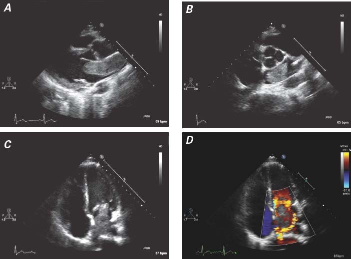
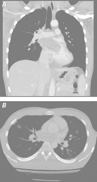
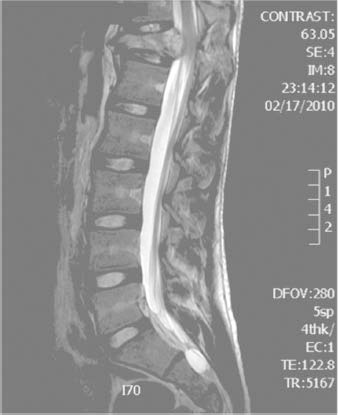
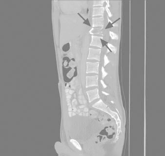
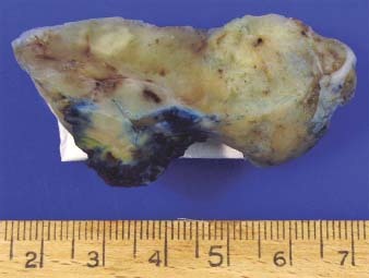
Soft-tissue sarcoma is the most prevalent primary malignant cardiac tumor. This sarcoma usually presents with cardiac manifestations secondary to local obstruction or arrhythmias; very rarely does it present with initial symptoms of distant metastasis. We discuss the unusual case of an 18-year-old man who emergently presented with acute-on-chronic back pain. Imaging revealed a lesion on the 12th thoracic vertebra and a large mass arising from the left atrium. The cardiac mass was resected, and immunohistochemical analysis revealed it to be a pleomorphic sarcoma that had metastasized to the spine. The patient died 2 years later of diffuse metastases. In addition to the patient's case, we discuss the nature and treatment of cardiac sarcoma.
Keywords: Bone neoplasms/secondary; heart neoplasms/diagnosis/pathology/surgery; sarcoma/complications/diagnosis/surgery.
Figures

References
-
- Molina JE, Edwards JE, Ward HB. Primary cardiac tumors: experience at the University of Minnesota. Thorac Cardiovasc Surg 1990;38 Suppl 2:183–91. - PubMed
-
- Takach TJ, Reul GJ, Ott DA, Cooley DA. Primary cardiac tumors in infants and children: immediate and long-term operative results. Ann Thorac Surg 1996;62(2):559–64. - PubMed
-
- Herhusky MJ, Gregg SB, Virmani R, Chun PK, Bender H, Gray GF Jr. Cardiac sarcomas presenting as metastatic disease. Arch Pathol Lab Med 1985;109(10):943–5. - PubMed
Publication types
MeSH terms
Substances
LinkOut - more resources
Full Text Sources
Medical