. 2013 Aug;2(4):241-9.
doi: 10.1089/biores.2013.0021.

Immobilization of Cell-Adhesive Laminin Peptides in Degradable PEGDA Hydrogels Influences Endothelial Cell Tubulogenesis

Affiliations

Immobilization of Cell-Adhesive Laminin Peptides in Degradable PEGDA Hydrogels Influences Endothelial Cell Tubulogenesis

Saniya Ali et al. Biores Open Access. 2013 Aug.

Abstract

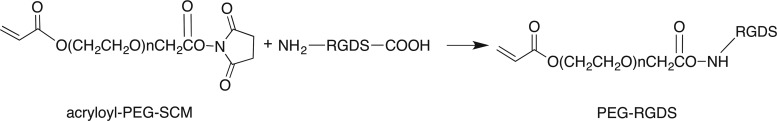
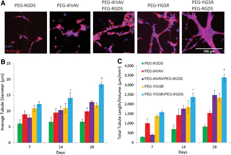
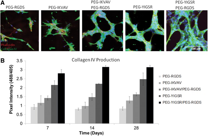
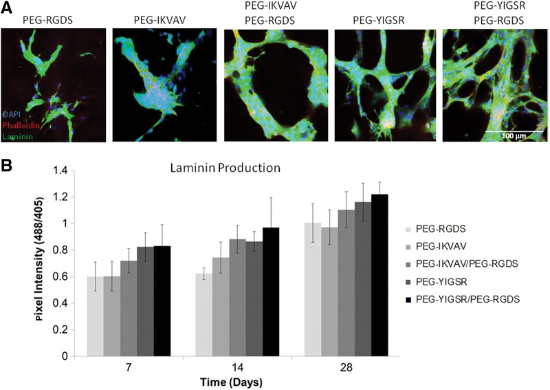
Attachment, spreading, and organization of endothelial cells into tubule networks are mediated by interactions between cells in the extracellular microenvironment. Laminins are key extracellular matrix components and regulators of cell adhesion, migration, and proliferation. In this study, laminin-derived peptides were conjugated to poly(ethylene glycol) (PEG) monoacrylate and covalently incorporated into degradable PEG diacrylate (PEGDA) hydrogels to investigate the influence of these peptides on endothelial cellular adhesion and function in organizing into tubule networks. Degradable PEGDA hydrogels were synthesized by incorporating a matrix metalloproteinase (MMP)-sensitive peptide, GGGPQGIWGQGK (abbreviated PQ), into the polymer backbone. The secretion of MMP-2 and MMP-9 by endothelial cells promotes polymer degradation and consequently cell migration. We demonstrate the formation of extensive networks of tubule-like structures by encapsulated human umbilical vein endothelial cells in hydrogels with immobilized synthetic peptides. The resulting structures were stabilized by pericyte precursor cells (10T1/2s) in vitro. During tubule formation and stabilization, extracellular matrix proteins such as collagen IV and laminin were deposited. Tubules formed in the matrix of metalloproteinase sensitive hydrogels were visualized from 7 days to 4 weeks in response to different combination of peptides. Moreover, hydrogels functionalized with laminin peptides and transplanted in a mouse cornea supported the ingrowth and attachment of endothelial cells to the hydrogel during angiogenesis. Results of this study illustrate the use of laminin-derived peptides as potential candidates for modification of biomaterials to support angiogenesis.

Keywords: angiogenesis; biomaterials; extracellular matrix; peptides; tissue engineering.


Figures

FIG. 1.
FIG. 1.
The cell-adhesive peptide, RGDS, was conjugated to acryloyl-PEG-SCM (3400 Da) to form PEG-RGDS. Similar reactions were used to conjugate the additional laminin peptides, yielding PEG-IKVAV and PEG-YIGSR. PEG, poly(ethylene glycol); SCM, succinimidyl carboxymethyl.
FIG. 2.
FIG. 2.
Images of cells in modified hydrogels (A) were taken after 7 days in culture. To quantify the resulting tubule formation in vitro, the average tubule diameter (B) and total tubule length per field of view (C) were calculated. *Significantly different from all other groups for time point 14 days in culture (p<0.01). xSignificantly different from all other groups at time point 28 days in culture (p<0.01). Supplementary Fig. S1 shows all individual significant differences between laminin peptide groups and their effect on average tubule diameter and total tubule length per volume.
FIG. 3.
FIG. 3.
The amount of extracellular matrix (ECM) proteins produced by tubules was dependent on the type of peptide presented to encapsulated cells. Images of cells in modified hydrogels (A) were taken after 14 days in culture (B). Laminin deposition was quantified and was found to correlate to the growth of tubules. Increase in laminin production (shown in green) by cells was observed as the time in culture was prolonged from 7 days to 28 days. Refer to Supplementary Fig. S2 for individual significant differences between laminin peptide groups and their effect on collagen IV production.
FIG. 4.
FIG. 4.
The amount of ECM proteins produced by tubules was dependent on the type of peptide presented to encapsulated cells. Images of cells in modified hydrogels (A) were taken after 14 days in culture (B). Collagen IV deposition shown in green was quantified and found to correspond to areas of high tubule formation. This result is noticeable for all time points up to 28 days for the PEG-YIGSR/PEG-RGDS combination. Refer to Supplementary Fig. S2 for individual significant differences between laminin peptide groups and their effect on laminin production.
FIG. 5.
FIG. 5.
As visualized by the mCherry fluorescence of mouse endothelial cells (A), the presence of PEG-YIGSR and PEG-RGDS induced a significant difference in vessel density (B), fractal dimension (C), lacunarity (D), vessel diameter (E), and branch points (F). *Significantly different from PEG-YIGSR and PEG-IKVAV (p<0.01). xSignificantly different from PEG-YIGSR and PEG-RGDS (p<0.05). #Significantly different from PEG-RGDS (p<0.05). ¥Significantly different from all other groups (p<0.05). Scale bar=50 μm. Significantly different from all other groups (p<0.01). +Significantly different from PEG-YIGSR and PEG-IKVAV (p<0.05).

References

    1. Atala A. Bauer SB. Soker S. Yoo JJ. Retik AB. Tissue-engineered autologous bladders for patients needing cystoplasty. Lancet. 2006;367:1241–1246. - PubMed
    1. Jain RK. Au P. Tam J. Duda DG. Fukumura D. Engineering vascularized tissue. Nat Biotechnol. 2005;23:821–823. - PubMed
    1. Francis ME. Uriel S. Brey EM. Endothelial cell-matrix interactions in neovascularization. Tissue Eng Part B Rev. 2008;14:19–32. - PubMed
    1. Nicosia RF. Madri JA. The microvascular extracellular matrix: developmental changes during angiogenesis in the aortic ring-plasma clot model. Am J Pathol. 1987;128:78–90. - PMC - PubMed
    1. Miner JH. Laminins and their roles in mammals. Microsc Res Tech. 2008;71:349–356. - PubMed