

Characterizing the normal proteome of human ciliary body
- PMID: 23914977
- PMCID: PMC3750387
- DOI: 10.1186/1559-0275-10-9
Characterizing the normal proteome of human ciliary body
Abstract
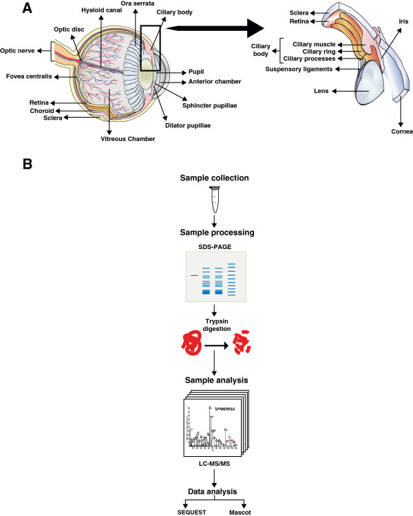
Background: The ciliary body is the circumferential muscular tissue located just behind the iris in the anterior chamber of the eye. It plays a pivotal role in the production of aqueous humor, maintenance of the lens zonules and accommodation by changing the shape of the crystalline lens. The ciliary body is the major target of drugs against glaucoma as its inhibition leads to a drop in intraocular pressure. A molecular study of the ciliary body could provide a better understanding about the pathophysiological processes that occur in glaucoma. Thus far, no large-scale proteomic investigation has been reported for the human ciliary body.
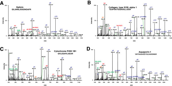
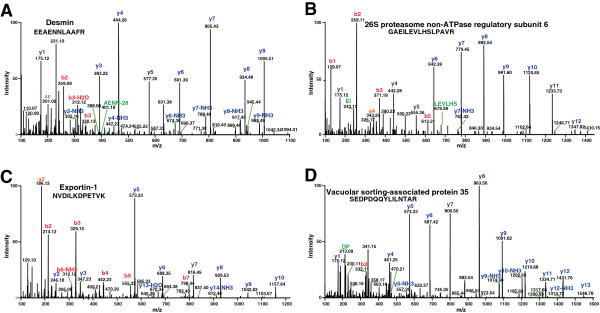
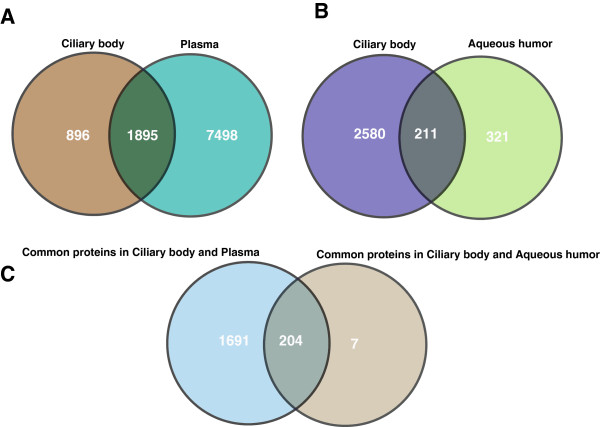
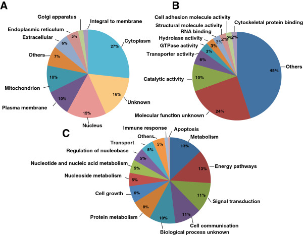
Results: In this study, we have carried out an in-depth LC-MS/MS-based proteomic analysis of normal human ciliary body and have identified 2,815 proteins. We identified a number of proteins that were previously not described in the ciliary body including importin 5 (IPO5), atlastin-2 (ATL2), B-cell receptor associated protein 29 (BCAP29), basigin (BSG), calpain-1 (CAPN1), copine 6 (CPNE6), fibulin 1 (FBLN1) and galectin 1 (LGALS1). We compared the plasma proteome with the ciliary body proteome and found that the large majority of proteins in the ciliary body were also detectable in the plasma while 896 proteins were unique to the ciliary body. We also classified proteins using pathway enrichment analysis and found most of proteins associated with ubiquitin pathway, EIF2 signaling, glycolysis and gluconeogenesis.
Conclusions: More than 95% of the identified proteins have not been previously described in the ciliary body proteome. This is the largest catalogue of proteins reported thus far in the ciliary body that should provide new insights into our understanding of the factors involved in maintaining the secretion of aqueous humor. The identification of these proteins will aid in understanding various eye diseases of the anterior segment such as glaucoma and presbyopia.
Figures
References
-
- Kaufman PL, Alm A, Adler FH. Adler's physiology of the eye: clinical application. 10. St. Louis: Mosby; 2003.
-
- Alm A, Bill A. Ocular and optic nerve blood flow at normal and increased intraocular pressures in monkeys (Macaca irus): a study with radioactively labelled microspheres including flow determinations in brain and some other tissues. Exp Eye Res. 1973;10:15–29. doi: 10.1016/0014-4835(73)90185-1. - DOI - PubMed
LinkOut - more resources
Full Text Sources
Other Literature Sources
Research Materials
Miscellaneous