. 2013 Aug 6:10:100.
doi: 10.1186/1742-2094-10-100.

Hemoglobin induces inflammation after preterm intraventricular hemorrhage by methemoglobin formation

Affiliations

Hemoglobin induces inflammation after preterm intraventricular hemorrhage by methemoglobin formation

Magnus Gram et al. J Neuroinflammation. .

Abstract

Background: Cerebral intraventricular hemorrhage (IVH) is a major cause of severe neurodevelopmental impairment in preterm infants. To date, no therapy is available that prevents infants from developing serious neurological disability following IVH. Thus, to develop treatment strategies for IVH, it is essential to characterize the initial sequence of molecular events that leads to brain damage. In this study, we investigated extracellular hemoglobin (Hb) as a causal initiator of inflammation in preterm IVH.

Methods: Using a preterm rabbit pup model, we investigated the molecular mechanisms and events following IVH. We also characterized the concentrations of cell-free Hb metabolites and pro-inflammatory mediators in the cerebrospinal fluid (CSF) of preterm human infants and rabbit pups. Finally, Hb metabolites were evaluated as causal initiators of inflammation in primary rabbit astrocyte cell cultures.

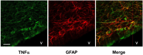
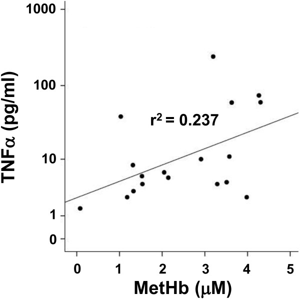
Results: Following IVH in preterm rabbit pups, the intraventricular CSF concentration of cell-free methemoglobin (metHb) increased from 24 to 72 hours and was strongly correlated with the concentration of TNFα at 72 hours (r2 = 0.896, P <0.001). Also, the mRNA expression of TNFα, IL-1β, and Toll-like receptor-4 and TNFα protein levels were significantly increased in periventricular tissue at 72 hours, which was accompanied by extensive astrocyte activation (that is, glial fibrillary acidic protein (GFAP)staining). Furthermore, exposure of primary rabbit astrocyte cell cultures to metHb caused a dose-dependent increase in TNFα mRNA and protein levels, which was not observed following exposure to oxyhemoglobin (oxyHb) or hemin. Finally, a positive correlation (r2 = 0.237, P <0.03) between metHb and TNFα concentrations was observed in the CSF of preterm human infants following IVH.

Conclusions: Following preterm IVH, increased metHb formation in the intraventricular space induces expression of pro-inflammatory cytokines. Thus, the formation of metHb might be a crucial initial event in the development of brain damage following preterm IVH. Accordingly, removal, scavenging, or neutralization of Hb could present a therapeutic opportunity and plausible approach to decreasing the damage in the immature brain following preterm IVH.


Figures

Figure 1
Figure 1
High-frequency ultrasound of normal brain and cerebral IVH. Coronal images obtained by high-frequency ultrasound displaying a normal brain with no IVH and cerebral IVH at six hours of age in preterm rabbit pups. Vertical ruler indicates 10 mm. IVH, intraventricular hemorrhage.
Figure 2
Figure 2
Hb metabolites and TNFα in CSF from rabbit pups following IVH. OxyHb (A), metHb (B), and TNFα (D) were quantified in intraventricular CSF at 24 (n = 6), 48 (n = 6), and 72 (n = 10) hours, as described in the Methods section, and the ratio of oxyHb/metHb was calculated (C). Horizontal lines depict median values. Median values of metHb and TNFα were increased at 72 hours as compared to corresponding values at 24 and 48 hours (P <0.01, Mann–Whitney U). The correlation between TNFα and metHb (E) at 72 hours was determined by linear regression analysis (r2 = 0.896, P <0.001). CSF, cerebrospinal fluid; Hb, hemoglobin; IVH, intraventricular hemorrhage; metHb, methemoglobin; oxyHb, oxyhemoglobin.
Figure 3
Figure 3
mRNA expression in periventricular brain tissue. mRNA expression of TNFα (A), IL-1β (B), TLR-4 (C) and HO-1 (D) in periventricular brain tissue of preterm rabbit pups with IVH (dark shaded bars; n = 6 at 24 hours and n = 6 at 72 hours) and in control pups (white bars; n = 10 at 24 hours and n = 17 at 72 hours). Expression of mRNA was determined using real-time PCR, as described in the Methods section, and levels of TNFα, IL-1β, TLR-4 and HO-1, respectively, were normalized against those of GAPDH and are given as fold change. The fold-change values were calculated by normalizing against control samples from untreated animals. Results are presented as box plots displaying medians and 25th and 75th percentiles. Differences between IVH versus control at 24 and 72 hours, respectively, were analyzed using the Mann–Whitney U test. * P <0.05, ** <0.01, *** <0.001. GAPDH, glyceraldehyde-3-phosphate dehydrogenase; HO-1, heme oxygenase; IVH, intraventricular hemorrhage; TLR-4, Toll-like receptor-4.
Figure 4
Figure 4
TNFα protein in periventricular brain tissue. Determination of TNFα protein concentration in periventricular brain tissue of preterm rabbit pups with IVH (dark shaded bars; n = 6 at 24 hours and n = 6 at 72 hours) and in control pups (white bars; n = 6 at 24 hours and n = 6 at 72 hours), using ELISA, as described in the Methods section. Results are presented as box plots displaying medians and 25th and 75th percentiles. The difference between IVH versus control at 72 hours was analyzed using the Mann–Whitney U test. * P <0.05. IVH, intraventricular hemorrhage.
Figure 5
Figure 5
TNFα and GFAP immunofluorescence following IVH. Immunofluorescence of rabbit pup brain sections at 72 hours following IVH was performed as described in the Methods section. Sections were stained against TNFα (green, AF-488) and GFAP (red, Cy3) and displayed a co-staining for TNFα in GFAP-positive astrocytes. Scale bar, 20 μm. V, ventricle. GFAP, glial fibrillary acidic protein; IVH, intraventricular hemorrhage.
Figure 6
Figure 6
Hb metabolite–induced TNFα, IL-1β, and HO-1 mRNA expression in astrocyte cell cultures. mRNA expression of TNFα (A), IL-1β (B) and HO-1 (C) in primary rabbit astrocyte cell cultures exposed to oxyHb, metHb, cyan-Hb, and hemin for four hours at concentrations of 1 μM (white bars), 5 μM (light shaded bars) and 15 μM (dark shaded bars), was determined using real-time PCR, as described in the Methods section. The mRNA expression of TNFα, IL-1β and HO-1 was normalized against GAPDH and is given as fold change. The fold-change values were calculated by normalizing against control samples from untreated cells. Results are from triplicate experiments and presented as mean ± SEM. Differences between the respective exposures and control conditions were analyzed using Mann–Whitney U. * P <0.05, ** <0.01. GAPDH, glyceraldehyde-3-phosphate dehydrogenase; Hb, hemoglobin; HO-1, heme oxygenase; metHb, methemoglobin; oxyHb, oxyhemoglobin; SEM, standard error of the mean.
Figure 7
Figure 7
Hb metabolite–induced TNFα protein secretion in astrocyte cell cultures. Determination of TNFα protein concentration in culture medium of primary rabbit astrocyte cell cultures, exposed for one to four hours to oxyHb, cyan-Hb, metHb and hemin at 1, 5 and 15 μM, respectively, using ELISA, as described in the Methods section. Exposure to oxyHb (filled circles) and cyan-Hb (open circles) is illustrated in panel A and metHb (filled squares) and hemin (open squares) in panel B. Continuous line = 15 μM; dotted line = 5 μM; hatched line = 1 μM. Results are from triplicate experiments and are presented as mean ± SEM. MetHb at 1, 5 and 15 μM versus control, all P <0.01 (ANOVA for repeated measures). ANOVA, analysis of variance; Hb, hemoglobin; metHb, methemoglobin; oxyHb, oxyhemoglobin; SEM, standard error of the mean.
Figure 8
Figure 8
Fenton reaction–induced TLR-4, TNFα, IL-1β and HO-1 mRNA expression in astrocyte cell cultures. Primary rabbit astrocyte cell cultures were exposed to a mixture of (NH4)Fe(SO4)2, hydrogen peroxide and ascorbate (the Fenton reaction) for four hours at concentrations of 10 μM (NH4)Fe(SO4)2 + 100 μM ascorbate + 20 μM H2O2 (white bars) or 50 μM (NH4)Fe(SO4)2, 500 μM ascorbate + 100 μM H2O2 (shaded bars). mRNA expression of TLR-4 (A), TNFα (B), IL-1β (C) and HO-1 (D) was determined using real-time PCR, as described in the Methods section. The mRNA expression of TLR-4, TNFα, IL-1β and HO-1 was normalized against GAPDH and is given as fold change. The fold-change values were calculated by normalizing against control samples from untreated cells. Results are from triplicate experiments and presented as mean ± SEM. Differences between the respective exposures and control conditions were analyzed using Mann–Whitney U. *** P <0.001. GAPDH, glyceraldehyde-3-phosphate dehydrogenase; HO-1, heme oxygenase; SEM, standard error of the mean; TLR-4, Toll-like receptor-4.
Figure 9
Figure 9
Hb metabolites and TNFα in CSF from preterm infants following IVH. Levels of metHb and TNFα from CSF obtained in serial samples from four preterm infants with IVH were determined as described in the Methods section. The correlation between TNFα and metHb was determined by linear regression analysis (r2 = 0.237, P = 0.01). CSF, cerebrospinal fluid; Hb, hemoglobin; IVH, intraventricular hemorrhage; metHb, methemoglobin.

References

    1. Stoll BJ, Hansen NI, Bell EF, Shankaran S, Laptook AR, Walsh MC, Hale EC, Newman NS, Schibler K, Carlo WA, Kennedy KA, Poindexter BB, Finer NN, Ehrenkranz RA, Duara S, Sánchez PJ, O'Shea TM, Goldberg RN, Van Meurs KP, Faix RG, Phelps DL, Frantz ID 3rd, Watterberg KL, Saha S, Das A, Higgins RD. Eunice Kennedy Shriver National Institute of Child Health and Human Development Neonatal Research Network. Neonatal outcomes of extremely preterm infants from the NICHD Neonatal Research Network. Pediatrics. 2010;126:443–456. doi: 10.1542/peds.2009-2959. - DOI - PMC - PubMed
    1. Wilson-Costello D, Friedman H, Minich N, Fanaroff AA, Hack M. Improved survival rates with increased neurodevelopmental disability for extremely low birth weight infants in the 1990s. Pediatrics. 2005;115:997–1003. doi: 10.1542/peds.2004-0221. - DOI - PubMed
    1. Brouwer A, Groenendaal F, van Haastert IL, Rademaker K, Hanlo P, de Vries L. Neurodevelopmental outcome of preterm infants with severe intraventricular hemorrhage and therapy for post-hemorrhagic ventricular dilatation. J Pediatr. 2008;152:648–654. doi: 10.1016/j.jpeds.2007.10.005. - DOI - PubMed
    1. Murphy BP, Inder TE, Rooks V, Taylor GA, Anderson NJ, Mogridge N, Horwood LJ, Volpe JJ. Posthaemorrhagic ventricular dilatation in the premature infant: natural history and predictors of outcome. Arch Dis Child Fetal Neonatal Ed. 2002;87:F37–F41. doi: 10.1136/fn.87.1.F37. - DOI - PMC - PubMed
    1. Adams-Chapman I, Hansen NI, Stoll BJ, Higgins R. Neurodevelopmental outcome of extremely low birth weight infants with posthemorrhagic hydrocephalus requiring shunt insertion. Pediatrics. 2008;121:e1167–e1177. doi: 10.1542/peds.2007-0423. - DOI - PMC - PubMed

Publication types

MeSH terms

LinkOut - more resources