. 2013 Aug 1;6(1):10.
doi: 10.1186/2046-1682-6-10.

Molecular basis of HHQ biosynthesis: molecular dynamics simulations, enzyme kinetic and surface plasmon resonance studies

Affiliations

Molecular basis of HHQ biosynthesis: molecular dynamics simulations, enzyme kinetic and surface plasmon resonance studies

Anke Steinbach et al. BMC Biophys. .

Abstract

Background: PQS (PseudomonasQuinolone Signal) and its precursor HHQ are signal molecules of the P. aeruginosa quorum sensing system. They explicate their role in mammalian pathogenicity by binding to the receptor PqsR that induces virulence factor production and biofilm formation. The enzyme PqsD catalyses the biosynthesis of HHQ.

Results: Enzyme kinetic analysis and surface plasmon resonance (SPR) biosensor experiments were used to determine mechanism and substrate order of the biosynthesis. Comparative analysis led to the identification of domains involved in functionality of PqsD. A kinetic cycle was set up and molecular dynamics (MD) simulations were used to study the molecular bases of the kinetics of PqsD. Trajectory analysis, pocket volume measurements, binding energy estimations and decompositions ensured insights into the binding mode of the substrates anthraniloyl-CoA and β-ketodecanoic acid.

Conclusions: Enzyme kinetics and SPR experiments hint at a ping-pong mechanism for PqsD with ACoA as first substrate. Trajectory analysis of different PqsD complexes evidenced ligand-dependent induced-fit motions affecting the modified ACoA funnel access to the exposure of a secondary channel. A tunnel-network is formed in which Ser317 plays an important role by binding to both substrates. Mutagenesis experiments resulting in the inactive S317F mutant confirmed the importance of this residue. Two binding modes for β-ketodecanoic acid were identified with distinct catalytic mechanism preferences.


Figures

Figure 1
Figure 1
Ping-pong kinetic mechanism of PqsD in HHQ biosynthesis. A) Lineweaver-Burk Plot and B) Hanes-Woolf Plot representing the activity of PqsD as a function of ACoA concentration in the presence of 60 (diamonds), 120 (black circles), 240 (triangles), 480 (open squares) and 1000 μM (open circles) ßK. All data are mean values of four replicates. C) Kinetic parameters of PqsD. D) “Real-time” PqsD kinetics by SPR experiments: 1. ACoA injection; 2. buffer run - response units remain high due to anthranilate-Cys112; 3. βK injection; 4. Buffer run – response line goes to zero due to HHQ formation and restored apo PqsD. E) Schematic view of the putative ping-pong kinetic mechanism of the HHQ biosynthesis (E - PqsD, ECSJ - PqsD with Cys112-ligated anthranilate).
Figure 2
Figure 2
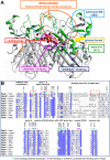
Functionally important domains of PqsD. A) 3D structure of the PqsD dimer with functionally significant domains and the central cavity. PqsD is rendered as cartoon with flexible regions coloured in green. Water molecules present in the central cavity are shown as red spheres. B) Multiple sequence alignment of PqsD and homologous KAS-III enzymes. Abbreviations: Paer – Pseudomonas aeruginosa, Bamb – Burkholderia ambifaria, Ecoli – Escherichia coli, Aael – Aquifex aeolicus, Tther – Thermus thermophilus, Saur – Staphylococcus aureus, Efaec – Enterococcus faecalis, Sliv – Streptomyces lividans, Mtub – Mycobacterium tuberculosis.
Figure 3
Figure 3
Residue-dependent RMS fluctuations of the apoform MD simulations with PqsD in its monomeric and dimer state. The fluctuations of the residues of PqsD in its monomer form (MD code A) are coloured in green, whereas for the PqsD in its dimer state (MD code B) chain A and B are shown in blue and red respectively. Domains putatively involved in the catalysis as identified by comparative analysis with homologous KAS-III enzymes are labelled with their abbreviations (dotted orange box indicates helix H14): adenosine binding site - aBS, palindromic “substrate-loop” - sL, helix H8-H9 loop - h8-9, hairpin-loop - hL, helix H12 - h12, “oxyanion-loop” – oL. Notably, also for all other MD simulations the largest RMS fluctuations were found in these areas (see also Additional file 1: Figure SI8).
Figure 4
Figure 4
Conformational changes at dimer interface in the dual-βK MD simulation E2b. Top view of the dimer interface at 30 ns with the secondary channel solvent-exposed (dotted yellow eclipse) and the βK molecules shown as green surfaces. The dashed green line indicates a cleft which exists between the two H11 helixes at 0 ns, but that disappears over the simulation run. The full yellow circles indicate additional grooves formed at the interface. The cyan arrows indicate the primary funnel accesses for both chains.
Figure 5
Figure 5
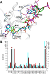
Binding mode of ACoA. A) Final poses of the single-ACoA simulation C1 (cyan; 37 ns) and of the dual-ACoA simulation C2 (green; 31 ns) are superimposed with the PDB structure 3H77 (ACoA orange - starting position; anthranilate-Cys112 in magenta – overlaps with ACoAC1). Phe32, Arg153, Asn154, Phe218, His257, Asn287 and Ser317 of the single-ACoA simulation at 37 ns are shown as dark cyan sticks. B) Decomposed energy contributions per residue (at least for one MD >0.5 kcal/mol) determined by MM-GBSA methods for the MD simulations C1 (cyan) and C2 (chain A – red; chain B – black).
Figure 6
Figure 6
Binding mode of βK in the secondary channel (MD simulations E2a and E2b). A) Superimposed snapshots of βK binding to PqsD at 0 and 30 ns. βK at 0 and 30 ns is shown as purple sticks and as green ball and sticks respectively. Important residues are shown as cyan lines at 30 ns and as orange lines at 0 ns. The red arrows indicate the conformational shift from the initial (yellow) to the final (blue) conformation of PqsD in the MD simulation E2b; Phe218 flips out in presence of βK. B) Schematic representation of βK in the secondary channel. Polar amino acids are illustrated in purple and hydrophobic amino acids in green circles. Hydrogen bonds and CH-pi interactions are shown as green arrows and dotted lines. C) Decomposed energy contributions per residue (at least for one MD >0.5 kcal/mol) determined by MM-GBSA methods for the MD simulations E2a (cyan) and E2b (chain A – red; chain B – black).
Figure 7
Figure 7
U-shaped channel network formed in presence of βK in the secondary channel. The U-shaped channel connects primary funnel access with the catalytic centre (large unfilled arrow), the secondary channel (thin arrow) and the dimer interface (large dotted arrows). In presence of βK a tunnel is formed between Cys112-bound anthranilate (CSJ) and the central cavity (dashed thin arrow) potentially used for water supply (needed for catalysis) from the central cavity to the oxyanion site close to P88′. βK is shown as green ball and sticks, whereas residues shown to interact with βK (CSJ, T195, F218, R145, D87′, P88′) as cyan sticks. For clarity the domains are labelled with their abbreviations. The solvent accessible surface is color-coded as follows: blue – positive electrostatic potential (+25 kcal/mol); red – negative electrostatic potential (−25 kcal/mol), white – neutral.
Figure 8
Figure 8
Binding modes of βK and putative catalytic mechanisms of βK-incorporation. A) βK is placed in the primary funnel (blue tube) as in the MD simulation E1: no space is left to host the thioester carrier CoA/ACP. B) The access of ACoA into the primary funnel is hindered with βK in the pose as in A. C) βK is in the secondary channel (violet box) as in the MD simulations E2a and E2b: in the primary funnel space is available to accommodate the thioester carrier CoA/ACP. D) ACoA can access the primary funnel with βK in this pose and transfer anthranilate to Cys112. E) The Claisen condensation fits best as mechanism for βK incorporation for βK in the primary funnel. F) The imine/enamine formation is the most plausible mechanism for βK incorporation when βK is in the secondary channel.
Figure 9
Figure 9
SPR sensograms of ACoA. When ACoA is injected with PqsD wild-type fixed on the chip (black) an increase in the response units is observed corresponding to the anthranilate transfer to Cys112. Differently, when S317F PqsD mutant (red) is present on the chip, no increase is observed.

Similar articles

Cited by

References

    1. Schuster M, Greenberg EP. A network of networks: Quorum-sensing gene regulation in Pseudomonas aeruginosa. Int J Med Microbiol. 2006;296:73–81. doi: 10.1016/j.ijmm.2006.01.036. - DOI - PubMed
    1. Williams P, Cámara M. Quorum sensing and environmental adaptation in Pseudomonas aeruginosa: a tale of regulatory networks and multifunctional signal molecules. Curr Opin Microbiol. 2009;12:182–191. doi: 10.1016/j.mib.2009.01.005. - DOI - PubMed
    1. Bjarnsholt T, Givskov M. The role of quorum sensing in the pathogenicity of the cunning aggressor Pseudomonas aeruginosa. Anal Bioanal Chem. 2007;387:409–414. doi: 10.1007/s00216-006-0774-x. - DOI - PubMed
    1. Jimenez PN, Koch G, Thompsona JA, Xaviera KB, Coolb RH, Quaxb WJ. The Multiple Signaling Systems Regulating Virulence in Pseudomonas aeruginosa. Microbiol Mol Biol Rev. 2012;76:46–65. doi: 10.1128/MMBR.05007-11. - DOI - PMC - PubMed
    1. Reen FJ, Mooij MJ, Holcombe LJ, McSweeney CM, McGlacken GP, Morrissey JP, O’Gara F. The Pseudomonas quinolone signal (PQS), and its precursor HHQ, modulate interspecies and interkingdom behaviour. FEMS Microbiol Ecol. 2011;77:413–428. doi: 10.1111/j.1574-6941.2011.01121.x. - DOI - PubMed

LinkOut - more resources